Greater tubercle
| Greater tubercle | |
|---|---|
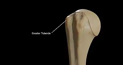
 Left humerus. Anterior view. (Greater tubercle visible at right.) | |
| Details | |
| Identifiers | |
| Latin | tuberculum majus humeri |
| TA98 | A02.4.04.005 |
| TA2 | 1184 |
| FMA | 23390 |
| Anatomical terms of bone | |
The greater tubercle of the humerus is situated lateral to the head of the humerus and posterolateral to the lesser tubercle.
Structure

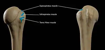
The upper surface of the greater tubercle is rounded, and marked by three flat impressions:
- the highest ("superior facet") gives insertion to the supraspinatus muscle.
- the middle ("middle facet") gives insertion to the infraspinatus muscle.
- the lowest ("inferior facet"), and the body of the bone for about 2.5 cm, gives insertion to the teres minor muscle.
The lateral surface of the greater tubercle is convex, rough, and continuous with the lateral surface of the body of the humerus. It can be described as having a cranial and a caudal part.[1]
Between the greater tubercle and the lesser tubercle is the bicipital groove (intertubercular sulcus).

Function
All three of the muscles that attach to the greater tubercle are part of the rotator cuff, a muscle group that stabilizes the shoulder joint. The greater tubercle therefore acts as a location for the transfer of forces from the rotator cuff muscles to the humerus.
The fourth muscle of the rotator cuff (subscapularis muscle) does not attach to the greater tubercle, but instead attaches to the lesser tubercle.
Clinical significance
The greater tubercle is usually the easiest part of the humerus to palpate.[2] It can be a useful surface landmark during surgery.[2]
Additional images
 Human arm bones diagram
References
![]() This article incorporates text in the public domain from page 209 of the 20th edition of Gray's Anatomy (1918)
This article incorporates text in the public domain from page 209 of the 20th edition of Gray's Anatomy (1918)
- ↑ Glass, Kati G.; Watkins, Jeffrey P. (2019-01-01), Auer, Jörg A.; Stick, John A.; Kümmerle, Jan M.; Prange, Timo (eds.), "Chapter 97 - Humerus", Equine Surgery (Fifth Edition), W.B. Saunders, pp. 1690–1699, ISBN 978-0-323-48420-6, retrieved 2021-01-08
- 1 2 Johnson, Kenneth A. (2014-01-01), Johnson, Kenneth A. (ed.), "Section 5 - The Forelimb", Piermattei's Atlas of Surgical Approaches to the Bones and Joints of the Dog and Cat (Fifth Edition), St. Louis: W.B. Saunders, pp. 158–309, ISBN 978-1-4377-1634-4, retrieved 2021-01-08
External links
- Anatomy figure: 03:02-09 at Human Anatomy Online, SUNY Downstate Medical Center
- Anatomy figure: 05:01-07 at Human Anatomy Online, SUNY Downstate Medical Center
- Anatomy figure: 10:02-13 at Human Anatomy Online, SUNY Downstate Medical Center
- aplab - BioWeb at University of Wisconsin System