Kiesselbach's plexus
| Kiesselbach's plexus | |
|---|---|
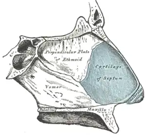
 The bones and cartilage of the nasal septum, viewed from right side. Kiesselbach's plexus (not labelled) is in the anterior inferior part of the nasal septum known as Little's area. | |
| Details | |
| Location | Little's area of nose |
| From | anterior ethmoidal artery, sphenopalatine artery, greater palatine artery, septal branch of superior labial artery, posterior ethmoidal artery |
| Supplies | nasal septum |
| Anatomical terminology | |
Kiesselbach's plexus is a vascular network of four or five arteries in the nose. It supplies the nasal septum. The arteries anastomose to form the plexus. It lies in the anterior inferior part of the septum known as Little's area, Kiesselbach's area, or Kiesselbach's triangle. It is a common site for nosebleeds.
Structure
Kiesselbach's plexus is an anastomosis of four or five arteries:
- the anterior ethmoidal artery, a branch of the ophthalmic artery.[1][2]
- the sphenopalatine artery, a terminal branch of the maxillary artery.[1][2]
- the greater palatine artery, a branch of the maxillary artery.[1][2]
- a septal branch of the superior labial artery, a branch of the facial artery.[1][2]
- a posterior ethmoidal artery, a branch of the ophthalmic artery.[1] There is contention as whether this is truly part of Kiesselbach's plexus. Most sources quote that it is not part of the plexus, but rather one of the blood supplies for the nasal septum itself.[2]
It runs vertically downwards just behind the columella, and crosses the floor of the nose. It joins the venous plexus on the lateral nasal wall.
Function
Kiesselbach's plexus supplies blood to the nasal septum.[2]
Clinical significance
Ninety percent of nosebleeds (epistaxis) occur in Kiesselbach's plexus.[3] It is exposed to the drying effect of inhaled air.[3] It can also be damaged by trauma from a finger nail (nose-picking), as it is fragile.[3][4] It is the usual site for nosebleeds in children and young adults.[3][5] A physician may use a nasal speculum to see that an anterior nosebleed comes from Kiesselbach's plexus.[6]
History
James Lawrence Little (1836–1885), an American surgeon, first described the area in detail in 1879. Little described the area as being "about half an inch ... from the lower edge of the middle of the column [septum]".[7]
Kiesselbach's plexus is named after Wilhelm Kiesselbach (1839–1902), a German otolaryngologist who published a paper on the area in 1884. The area may be called Little's area,[4] Kiesselbach's area, or Kiesselbach's triangle.
See also
References
- 1 2 3 4 5 Moore, Keith L. et al. (2014) Clinically Oriented Anatomy, 7th Ed, p.959
- 1 2 3 4 5 6 Drake, Richard L. (2005). Gray's anatomy for students. Wayne Vogl, Adam W. M. Mitchell, Henry Gray. Philadelphia: Elsevier / Churchill Livingstone. pp. 978–979. ISBN 0-443-06612-4. OCLC 55139039.
{{cite book}}: CS1 maint: date and year (link) - 1 2 3 4 Doyle, DE (Mar 1986). "Anterior epistaxis: a new nasal tampon for fast, effective control". The Laryngoscope. 96 (3): 279–81. doi:10.1288/00005537-198603000-00008. PMID 3951304.
- 1 2 Morgan, Daniel J.; Kellerman, Rick (1 March 2014). "Epistaxis: Evaluation and Treatment". Primary Care: Clinics in Office Practice. 41 (1): 63–73. doi:10.1016/j.pop.2013.10.007. ISSN 0095-4543.
- ↑ Dhingra. Diseases of Ear,Nose and Throat. Elsevier.
- ↑ Ando, Yuji; Iimura, Jiro; Arai, Satoshi; Arai, Chiaki; Komori, Manabu; Tsuyumu, Matsusato; Hama, Takanori; Shigeta, Yasushi; Hatano, Atsushi; Moriyama, Hiroshi (February 2014). "Risk factors for recurrent epistaxis: Importance of initial treatment". Auris Nasus Larynx. 41 (1): 41–45. doi:10.1016/j.anl.2013.05.004. ISSN 0385-8146.
- ↑ Little, James Lawrence (1879). "A hitherto undescribed lesion as a cause of epistaxis, with four cases". The Hospital Gazette. New York. 6 (1): 5–6.
External links
- Epistaxis - utmb.edu
- Nose Anatomy - emedicine.com
- Nasal Anatomy - fpnotebook.com