Percutaneous nephrostomy
| Percutaneous nephrostomy | |
|---|---|
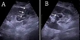 (A) Renal ultrasonograph of percutaneous nephrostomy tube placed through a calyx in the lower pole of a kidney with hydronephrosis. (B) The pigtail catheter is placed in the dilated calyx. The tube in (A) and the pigtail in (B) are marked with white arrows.[1] | |
| ICD-9-CM | 55.03 |
| OPS-301 code | 5-550 |
Percutaneous nephrostomy is an interventional radiology/surgical procedure in which the renal pelvis is punctured whilst using imaging as guidance. Images are obtained once an antegrade pyelogram (an injection of contrast), with a fine needle, has been performed. This contrast is used to show calcifications at the renal pelvis. A nephrostomy tube may then be placed to allow drainage.[2]
Medical uses
Diagnosis
Percutaneous nephrostomy is used in Whitaker test to differentiate recurrent obstruction or permanent dilatation after an operative surgery that corrects the cause of obstruction. This procedure is also used for antegrade pyelography to visualize the upper urinary tract system.[3]
Treatment
Percutaneous nephrostomy is also used to treat hydronephrosis caused by kidney stones, pregnancy, stricture of the urinary tract, urinary tract/cervical/prostate tumours. Besides, infections such as urosepsis and pyonephrosis can also be drained by nephrostomy tube insertion.[3] Percutaneous nephrostomy is also useful in divert urine away from diseased site to enhance healing. Examples of conditions that can be treated with such method are malignant/traumatic/inflammatory fistula, and haemorrhagic cystitis.[3]
Percutaneous nephrostomy is also used to provide access for chemotherapy/antibiotic/antifungal therapy, antegrade urethral stent placement, stone retrieval, and endopyelotomy (endoscopic surgery for the enlargement of the junction of renal pelvis and ureter).[3]
Technique
Informed consent from the patient should be taken before the procedure. Besides, intravenous access and antibiotics should be given half an hour before the procedure for those with underlying urinary infection.[3]
Then the subject lie down in prone position with the side to be operated on lie at the edge of the table. The area is then cleaned and draped. If the subject is unable to lie down prone due to underlying heart or lung problem, the supine position can also be used.[3]
An area for needle insertion is marked at the region with four sides, namely: upper margin are the borders of 11th and 12th ribs, lower margin is the iliac crest, medial margin is marked by the muscles beside the spine, and lateral margin is marked by the posterior axillary line.
The puncture area is the located using ultrasound. Local anaesthetic is then injected around the area and scalpel incision is made. An 18 G trocar needle is inserted into the puncture site. The operator can feel two "pop"s while inserting the needle, one is when passing through the renal capsule, and another one is when entering the collecting system. Then urine is aspirated from the collecting system and sent for culture. If the urine is clear, then dye is injected into the system for delineation of calyceal system. If the urine is turbid, dye should not be injected into the collecting system to prevent the spread of infection.[3]
Side effects
Among the side effects of percuteneous nephrosotomy are haematuria (blood in urine), pain, sepsis, injury to adjacent organs, extravasaction or urine, catheter dislodgement, and inability to remove nephrostomy tube due to crystallisation. Although pneumothorax and colonic injury are more common on subcostal needle insertion, these are rare complications.[3]
Blood in urine usually clears up after 48 to 72 hours. Bleeding longer than this period may signifies more serious bleeding complication. About 2 to 4% of percutaneous nephrostomy cases require blood transfusion.[4]
See also
References
- ↑ Content initially copied from: Hansen KL, Nielsen MB, Ewertsen C (December 2015). "Ultrasonography of the Kidney: A Pictorial Review". Diagnostics. 6 (1): 2. doi:10.3390/diagnostics6010002. PMC 4808817. PMID 26838799. (CC-BY 4.0)
- ↑ Longmore M, Wilkinson I, Turmezei T, Cheung CK (2007). Oxford Handbook of Clinical Medicine (7th ed.). Oxford University Press. p. 731. ISBN 978-0-19-856837-7.
- 1 2 3 4 5 6 7 8 Jairath A, Ganpule A, Desai M (2017-11-29). "Percutaneous nephrostomy step by step". Mini-invasive Surgery. 1: 180–185. doi:10.20517/2574-1225.2017.24. ISSN 2574-1225.
- ↑ Smith M, Rochon PJ, Ray CE (June 2012). "Traversing the Renal Pelvis during Percutaneous Nephrostomy Tube Placement ("Kidney Kabob")". Seminars in Interventional Radiology. 29 (2): 150–152. doi:10.1055/s-0032-1312578. PMC 3444877. PMID 23729987.