Splinter hemorrhage
| Splinter hemorrhage | |
|---|---|
 | |
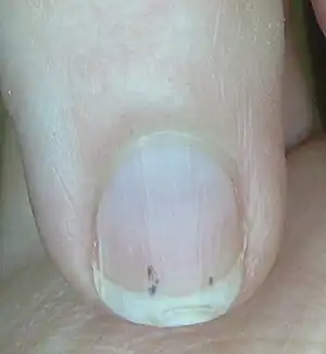
| Splinter hemorrhage on a fingernail of the little finger | |
| Differential diagnosis | subacute infective endocarditis, scleroderma, trichinosis, systemic lupus erythematosus (SLE), rheumatoid arthritis, psoriatic nails, antiphospholipid syndrome |
Splinter hemorrhages (or haemorrhages), are reddish longitudinal lines in a nail, frequently associated with endocarditis, systemic lupus erythematosus (SLE) and trichinosis.[1] They can occur on their own or be associated with trauma, nail tumor or some inflammatory skin conditions, when it may occur in multiple nails and be painful.[2] The lines are typically fine, do not blanche, may be red to black, measure around 1-to-3mm, and grow out with the nail.[2]
They occur when blood enters the nail ridges due to bleeding of small blood vessels in the nail bed.[2] Dermoscopy shows red-to-black lines that fade towards the nail tip.[2] Treatment depends on cause.[2]
Signs and symptoms
.jpg.webp) Splinter hemorrhage
Splinter hemorrhage.jpg.webp) Splinter hemorrhage
Splinter hemorrhage.jpg.webp) Splinter hemorrhage
Splinter hemorrhage.jpg.webp) Splinter hemorrhage
Splinter hemorrhage.jpg.webp) Splinter hemorrhage seen end on
Splinter hemorrhage seen end on Splinter hemorrhage under the microscope
Cause
Splinter hemorrhages are tiny blood clots that tend to run vertically under the nails. They are not specific to any particular condition, and can be associated with subacute infective endocarditis, scleroderma, trichinosis, systemic lupus erythematosus (SLE), rheumatoid arthritis, psoriatic nails,[3] antiphospholipid syndrome,[4]: 659 haematological malignancy, and trauma.[5] At first they are usually plum-colored, but then darken to brown or black in a couple of days. In certain conditions (in particular, infective endocarditis), clots can migrate from the affected heart valve and find their way into various parts of the body. If this happens in the finger, it can cause damage to the capillaries resulting in a splinter hemorrhage. There are a number of other causes for splinter hemorrhages. They could be due to hitting the nail (trauma), a sign of inflammation in blood vessels all around the body (systemic vasculitis), or they could be where a fragment of cholesterol has become lodged in the capillaries of the finger. Even if a person does have infective endocarditis, roughly one in 10 people have splinter hemorrhages.[6]
See also
References
- ↑ Johnstone, Ronald B. (2017). "2. Diagnostic clues and "need-to-know" items". Weedon's Skin Pathology Essentials (2nd ed.). Elsevier. p. 32. ISBN 978-0-7020-6830-0. Archived from the original on 2021-05-25. Retrieved 2022-04-15.
- 1 2 3 4 5 Dehavey, Florence; Richert, Bertrand (2021). "Nail in systemic disorders: main signs and clues". In Lipner, Shari (ed.). Nail Disorders: Diagnosis and Management, An Issue of Dermatologic Clinics. Philadelphia: Elsevier. p. 233. ISBN 978-0-323-70923-1.
- ↑ Li, Cindy (29 March 2011). "Nail Psoriasis: Overview of Nail Psoriasis". Medscape. Archived from the original on 17 April 2019. Retrieved 7 January 2012.
- ↑ Freedberg, et al. (2003). Fitzpatrick's Dermatology in General Medicine. (6th ed.). McGraw-Hill. ISBN 0-07-138076-0.
- ↑ Rapini, Ronald P.; Bolognia, Jean L.; Jorizzo, Joseph L. (2007). Dermatology: 2-Volume Set. St. Louis: Mosby. ISBN 978-1-4160-2999-1.
- ↑ Hoen, Bruno (April 11, 2013). "Infective Endocarditis". New England Journal of Medicine. 368 (15): 1425–33. doi:10.1056/NEJMcp1206782. PMID 23574121.