Sagittal suture
| Sagittal suture | |
|---|---|
 Human adult skull from above. | |
 Human adult skull from above. Sagittal suture labeled at center. | |
| Details | |
| Identifiers | |
| Latin | sutura sagittalis |
| TA98 | A03.1.02.003 |
| TA2 | 1576 |
| FMA | 52929 |
| Anatomical terminology | |
The sagittal suture, also known as the interparietal suture and the sutura interparietalis, is a dense, fibrous connective tissue joint between the two parietal bones of the skull. The term is derived from the Latin word sagitta, meaning arrow.
Structure
The sagittal suture is formed from the fibrous connective tissue joint between the two parietal bones of the skull.[1] It has a varied and irregular shape which arises during development.[1] The pattern is different between the inside and the outside.[1]
Two anatomical landmarks are found on the sagittal suture: the bregma, and the vertex of the skull. The bregma is formed by the intersection of the sagittal and coronal sutures. The vertex is the highest point on the skull and is often near the midpoint of the sagittal suture.
Development
At birth, the bones of the skull do not meet.[1] The gap that remains, which is approximately 5 mm wide, allows for the brain to continue to grow normally after birth.[1] The inner parts of the parietal bones fuse before the outer parts.[1]
Clinical significance
If certain bones of the skull grow too fast before birth, then "premature closure" of the sutures may occur.[2] This can cause craniosynostosis, which results in skull deformities.[2] Sagittal craniosynostosis is the most common form.[2]
If the sagittal suture closes early the skull becomes long, narrow, and wedge-shaped, a condition called scaphocephaly.
Society and culture
In forensic anthropology, the sagittal suture is one method used to date human remains. The suture begins to close at age twenty nine, starting at where it intersects at the lambdoid suture and working forward. By age thirty five, the suture is completely closed. This means that when inspecting a human skull, if the suture is still open, one can assume an age of less than twenty nine. Conversely, if the suture is completely formed, one can assume an age of greater than thirty five.
History
The term is derived from the Latin word sagitta, meaning arrow. The derivation of this term may be demonstrated by observing how the sagittal suture is notched posteriorly, like an arrow, by the lambdoid suture.
The sagittal suture is also known as the interparietal suture, the sutura interparietalis.
Additional images
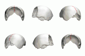
 Animation. Sagittal suture shown in red.
 Left and right parietal bone.
 Sagittal suture seen from inside.
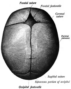
 Sagittal suture of a new-born child, seen from above.
 Sagittal suture of a new-born child.
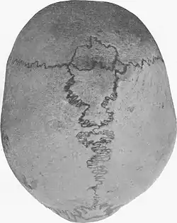
 Sagittal suture with wormian bones.
References
- 1 2 3 4 5 6 Oota, Yasuhito; Ono, Kenzo; Miyazima, Sasuke (2006-01-01). "3D modeling for sagittal suture". Physica A: Statistical Mechanics and Its Applications. 359: 538–546. Bibcode:2006PhyA..359..538O. doi:10.1016/j.physa.2005.05.095. ISSN 0378-4371.
- 1 2 3 "Sagittal craniosynostosis". www.gosh.nhs.uk. Retrieved 2021-01-04.
Bibliography
- "Sagittal suture", Stedman's Medical Dictionary, 27th ed. (2000).
- Moore, Keith L., and T.V.N. Persaud. The Developing Human: Clinically Oriented Embryology, 7th ed. (2003).
External links
| Wikimedia Commons has media related to Sagittal sutures. |
- Anatomy figure: 22:03-01 at Human Anatomy Online, SUNY Downstate Medical Center
- Anatomy image: skel/behind2 at Human Anatomy Lecture (Biology 129), Pennsylvania State University