ANO5
Anoctamin 5 (ANO5) is a protein that in humans is encoded by the ANO5 gene.
| ANO5 | |||||||||||||||||||||||||||||||||||||||||||||||||||
|---|---|---|---|---|---|---|---|---|---|---|---|---|---|---|---|---|---|---|---|---|---|---|---|---|---|---|---|---|---|---|---|---|---|---|---|---|---|---|---|---|---|---|---|---|---|---|---|---|---|---|---|
| Identifiers | |||||||||||||||||||||||||||||||||||||||||||||||||||
| Aliases | ANO5, GDD1, LGMD2L, TMEM16E, anoctamin 5, LGMDR12 | ||||||||||||||||||||||||||||||||||||||||||||||||||
| External IDs | OMIM: 608662 MGI: 3576659 HomoloGene: 100071 GeneCards: ANO5 | ||||||||||||||||||||||||||||||||||||||||||||||||||
| |||||||||||||||||||||||||||||||||||||||||||||||||||
| |||||||||||||||||||||||||||||||||||||||||||||||||||
| |||||||||||||||||||||||||||||||||||||||||||||||||||
| |||||||||||||||||||||||||||||||||||||||||||||||||||
| |||||||||||||||||||||||||||||||||||||||||||||||||||
| Wikidata | |||||||||||||||||||||||||||||||||||||||||||||||||||
| |||||||||||||||||||||||||||||||||||||||||||||||||||
Function
The ANO5 gene provides instructions for making a protein called anoctamin-5. While the specific function of this protein is not well understood, it belongs to a family of proteins, called anoctamins, that act as chloride channels. Chloride channels, which transport negatively charged chlorine atoms (chloride ions) in and out of cells, play a key role in a cell's ability to generate and transmit electrical signals. Most anoctamin proteins function as chloride channels that are turned on (activated) in the presence of positively charged calcium atoms (calcium ions); these channels are known as calcium-activated chloride channels. The mechanism for this calcium activation is unclear. Anoctamin proteins are also involved in maintaining the membrane that surrounds cells and repairing the membrane if damaged.[5]
The anoctamin-5 protein is most abundant in muscles used for movement (skeletal muscles). For the body to move normally, skeletal muscles must tense (contract) and relax in a coordinated way. The regulation of chloride flow within muscle cells plays a role in controlling muscle contraction and relaxation.[5]
The anoctamin-5 protein is also found in other cells including heart (cardiac) muscle cells and bone cells. The anoctamin-5 protein may be important for the development of muscle and bone before birth.[5]
Diseases associated with ANO5 mutations
- Gnathodiaphyseal dysplasia (GDD), a rare skeletal syndrome.[6]
- Limb Girdle Muscular Dystrophy 2L (LGMD2L, Autosomal Recessive 12)[6][7] and Miyoshi Muscular Dystrophy 3 (MMD3).[6] These forms of muscular dystrophy are inherited in an autosomal recessive pattern. To be affected, a person must have mutations on both copies of the gene. Males and females are equally likely to be affected.
Typical Symptoms
GDD causes bone fragility, sclerosis of tubular bones, and cemento-osseous lesions of the jawbone. Patients also experience frequent bone fractures.[6]
Clinically, LGMD2L and MMD3 were considered different diseases before ANO5 was identified as the responsible gene; LGMD was used to describe initial weakness in proximal muscles (hip and shoulder girdles) while MMD described initial weakness in the distal muscles of the lower limbs.[6]
Other names for this gene
- ANO5_HUMAN
- anoctamin-5
- GDD1
- gnathodiaphyseal dysplasia 1 protein
- integral membrane protein GDD1
- LGMD2L
- TMEM16E
- transmembrane protein 16E[5]
Chromosal location
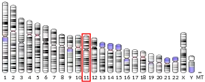
- Cytogenetic location: 11p14.3, which is the short (p) arm of chromosome 11 at position 14.3
- Molecular location: base pairs 22,192,485 to 22,283,367 on chromosome 11 (Homo sapiens Annotation Release 109, GRCh38.p12) (NCBI)
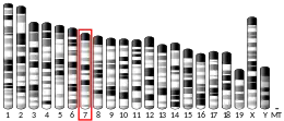
Credit: Genome Decoration Page/NCBI
References
- GRCh38: Ensembl release 89: ENSG00000171714 - Ensembl, May 2017
- GRCm38: Ensembl release 89: ENSMUSG00000055489 - Ensembl, May 2017
- "Human PubMed Reference:". National Center for Biotechnology Information, U.S. National Library of Medicine.
- "Mouse PubMed Reference:". National Center for Biotechnology Information, U.S. National Library of Medicine.
- "ANO5 gene". Genetics Home Reference. US National Library of Medicine. Retrieved 24 July 2018.
 This article incorporates text from this source, which is in the public domain.
 - "UniProt". www.uniprot.org. Retrieved 2023-09-20.
- "Autosomal recessive limb-girdle muscular dystrophy type 2L (Concept Id: C1969785) - MedGen - NCBI". www.ncbi.nlm.nih.gov. Retrieved 2023-09-20.
Further reading
- Penttilä S, Palmio J, Udd B (November 2012). "ANO5-Related Muscle Diseases". In Adam MP, Ardinger HH, Pagon RA, Wallace SE, Bean LJ, Stephens K, Amemiya A, Penttilä S, Palmio J, Udd B (eds.). SourceGeneReviews [Internet]. Seattle (WA): University of Washington, Seattle. PMID 23193613.
- "What is LGMD2L?". LGMD2L foundation.
- Online Mendelian Inheritance in Man (OMIM): ANOCTAMIN 5; ANO5 - 608662
- Bolduc V, Marlow G, Boycott KM, Saleki K, Inoue H, Kroon J, et al. (February 2010). "Recessive mutations in the putative calcium-activated chloride channel Anoctamin 5 cause proximal LGMD2L and distal MMD3 muscular dystrophies". American Journal of Human Genetics. 86 (2): 213–21. doi:10.1016/j.ajhg.2009.12.013. PMC 2820170. PMID 20096397.
- Jarmula A, Lusakowska A, Fichna JP, Topolewska M, Macias A, Johnson K, et al. (August 2019). "ANO5 mutations in the Polish limb girdle muscular dystrophy patients: Effects on the protein structure". Scientific Reports. 9 (1): 11533. Bibcode:2019NatSR...911533J. doi:10.1038/s41598-019-47849-3. PMC 6687736. PMID 31395899.


