Gastralia
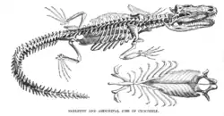
Gastralia (singular gastralium) are dermal bones found in the ventral body wall of modern crocodilians and tuatara, and many prehistoric tetrapods. They are found between the sternum and pelvis, and do not articulate with the vertebrae. In these reptiles, gastralia provide support for the abdomen and attachment sites for abdominal muscles.


The possession of gastralia may be ancestral for Tetrapoda and were possibly derived from the ventral scales found in animals like rhipidistians, labyrinthodonts, and Acanthostega, and may be related to ventral elements of turtle plastrons.[1][2] Similar, but not homologous cartilagenous elements, are found in the ventral body walls of lizards and anurans. These structures have been referred to as inscriptional ribs,[2] based on their alleged association with the inscriptiones tendinae (the tendons that form the six pack in humans). However, the terminology for these gastral-like structures remains confused. Both types, along with sternal ribs (ossified costal cartilages), have been referred to as abdominal ribs, a term with limited usefulness that should be avoided.[2] Gastralia are also present in a variety of extinct animals, including theropod and prosauropod dinosaurs, pterosaurs, plesiosaurs, choristoderes and some primitive pelycosaurs. In dinosaurs, the elements articulate with each other in a sort of zig-zag along the midline and may have aided in respiration.[2] Gastralia are known to be present in primitive ornithischian and sauropodomorph dinosaurs. However gastralia are only known from heterodontosaurid ornithschians, and gastralia are lost in eusauropodan sauropods.[3][4]
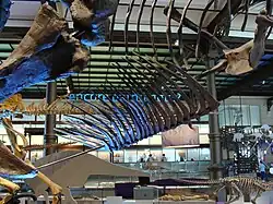
Discoveries about how the gastralia fit together in the skeleton of Sue the T. rex have led to an understanding that Tyrannosaurus bodies were more barrel-chested – and heavier – than previously thought.[5]
Pathology
The Allosaurus fragilis specimen USNM 8367 contained several gastralia which preserve evidence of healed fractures near their middle. Some of the fractures were poorly healed and "formed pseudoarthroses." An apparent subadult male Allosaurus fragilis was reported by Laws to have extensive pathologies. The possible subadult A. jimmadseni[6] specimen MOR 693 also had pathological gastralia.[7] The left scapula and fibula of an Allosaurus fragilis specimen catalogued as USNM 4734 are both pathological, both probably due to healed fractures.[8]
The holotype of Neovenator salerii had many pathologies, including pseudoarthrotic gastralia and a deviation to the right of the third and fourth neural spines of the neck vertebrae.[8]
An immature dromaeosaurid specimen (which hadn't been described in the scientific literature as of 2001) from Tugrugeen Shireh was observed to have a "bifurcated" gastralium.[8]
In the Gorgosaurus libratus holotype (NMC 2120) the 13th and 14th gastralia have healed fractures. Another G. libratus specimen catalogued as TMP94.12.602 bears multiple pathologies, including a pseudoarthortic gastralium.[8]
The unidentified tyrannosaurid specimen TMP97.12.229 had a fractured and healed gastralium.[8]
References
- Kardong KV (2002). Vertebrates: Comparative Anatomy, Function, Evolution (3rd ed.). New York: McGraw-Hill. pp. 291–293. ISBN 0-07-290956-0.
- Claessens LP (March 2004). "Dinosaur gastralia: origin, morphology, and function" (PDF). Journal of Vertebrate Paleontology. 24 (1): 89–106. doi:10.1671/A1116-8. S2CID 53318713.
- Tschopp E, Mateus O (March 2013). "Clavicles, interclavicles, gastralia, and sternal ribs in sauropod dinosaurs: new reports from diplodocidae and their morphological, functional and evolutionary implications". Journal of Anatomy. 222 (3): 321–40. doi:10.1111/joa.12012. PMC 3582252. PMID 23190365.
- Radermacher VJ, Fernandez V, Schachner ER, Butler RJ, Bordy EM, Naylor Hudgins M, et al. (July 2021). Long JA, Perry GH, Spencer M (eds.). "A new Heterodontosaurus specimen elucidates the unique ventilatory macroevolution of ornithischian dinosaurs". eLife. 10: e66036. doi:10.7554/eLife.66036. PMC 8260226. PMID 34225841.
- "A Fresh Science Makeover for SUE". 30 November 2018. Retrieved 17 December 2021.
- Chure DJ, Loewen MA (2020). "Cranial anatomy of Allosaurus jimmadseni, a new species from the lower part of the Morrison Formation (Upper Jurassic) of Western North America". PeerJ. 8: e7803. doi:10.7717/peerj.7803. PMC 6984342. PMID 32002317.
- Hanna RR (March 2002). "Multiple injury and infection in a sub-adult theropod dinosaur Allosaurus fragilis with comparisons to allosaur pathology in the Cleveland-Lloyd Dinosaur Quarry collection". Journal of Vertebrate Paleontology. 22 (1): 76–90. doi:10.1671/0272-4634(2002)022[0076:MIAIIA]2.0.CO;2. S2CID 85654858.
- Molnar RE (2001). "Theropod paleopathology: a literature survey". In Tanke DH, Carpenter K (eds.). Mesozoic Vertebrate Life. Indiana University Press. pp. 337–363.