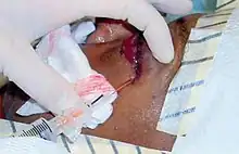
Canthotomy
Canthotomy (also called lateral canthotomy and canthotomy with cantholysis) is a surgical procedure where the lateral canthus, or corner, of the eye is cut to relieve the fluid pressure inside or behind the eye, known as intraocular pressure (IOC).[1] The procedure is typically done in emergency situations when the intraocular pressure becomes too high, which can damage the optic nerve and lead to blindness if left untreated.[2]
| Canthotomy | |
|---|---|
 Eye anatomy demonstrating the medial canthus | |
| Pronunciation | kăn-thŏt′ə-mē |
| Other names | Lateral canthotomy, canthotomy with cantholysis |
| Specialty | Ophthalmology and emergency medicine |
| Complications | Iatrogenic globe injury, bleeding, infection |
The most common cause of elevated intraocular pressure is orbital compartment syndrome (OCS) caused by trauma, retrobulbar hemorrhage, infections, tumors, or prolonged hypoxemia.[3] Absolute contraindications to canthotomy include globe rupture. Complications include bleeding, infections, cosmetic deformities, and functional impairment of eyelids.[3] Lateral canthotomy further specifies that the lateral canthus is being cut. Canthotomy with cantholysis includes cutting the lateral palpebral ligament, also known as the canthal tendon.
History
The first case of orbital compartment syndrome causing monocular blindness was published in 1950 due to a complication of a zygomatic fracture repair.[4] In 1953 the first surgical orbital decompression was performed. Two incisions below and above the external canthus were made and surgical drains were put in place.[5] In 1990 the first lateral canthotomy procedure as presently performed was completed.[6] In 1994 lateral canthotomy was first published in a review of procedures that emergency physicians can perform. [7] Today a canthotomy is almost always performed with cantholysis of the inferior canthal tendon as this provides the best decompression of intraocular pressure.[8]
Indications
A canthotomy is often used as a last resort to decompress orbital compartment syndrome. Orbital compartment syndrome can be caused by trauma, infections, tumors, retrobulbar hemorrhage, or prolonged hypoxemia.[9] Orbital compartment syndrome can be recognized by elevated intraocular pressure, globe compressibility, afferent pupillary defect, proptosis, decreased visual acuity, and decreased extraocular muscle movements.
Studies in animals have demonstrated irreversible vision loss within 90 to 120 minutes, further indicating the emergent nature of this procedure. [8]
In an unconscious patient who is unable to comply with a physical exam, an intraocular pressure greater than 40 indicates emergent canthotomy.[2]
Contraindications

The foremost absolute contraindication to canthotomy is globe rupture, sometimes referred to as an open globe injury.[9] Globe rupture can be recognized by these symptoms or physical exam features:
- Irregular-shaped pupil or iris
- Subconjunctival hemorrhage
- Enophthalmos
- Conjunctival or scleral tear
Due to the emergent nature of this procedure and the possibility of restoring or preventing vision loss, globe rupture is the only absolute contraindication.
Complications

Due to portions of the procedure having poor visualization of anatomical structures, and the overall rarity and difficulty of the procedure, iatrogenic globe injury is an immediate complication that can occur. Other complications include infections, bleeding, cosmetic deformities, and functional impairment of eyelids.[3]
Alternatives
Due to the infrequency and difficulty of canthotomy, emergency medicine physicians defer more than 50 percent of canthotomies to a consulting physician,[10] which in turn can increase time to treatment. In an effort to decrease difficulty and improve patient outcomes, vertical lid split or paracanthal "one-snip" procedures have been studied. This is performed by making a full-thickness vertical incision a few millimeters medial from the lateral canthus in both the upper and lower eyelids.[11]
References
- Nagelhout, John J.; Plaus, Karen (2009). "Chapter 40. Anesthesia For Ophthalmic Procedures". Nurse Anesthesia. Elsevier Health Sciences. p. 963. ISBN 9780323081016. Retrieved March 24, 2023 – via Google Books.
Canthotomy is a procedure performed to increase the orbital space by cutting the lateral canthus. This procedure reduces the orbital pressure that results from a retrobulbar hemorrhage.
- McInnes, Gord; Howes, Daniel W. (January 2002). "Lateral canthotomy and cantholysis: a simple, vision-saving procedure". CJEM. 4 (1): 49–52. doi:10.1017/s1481803500006060. ISSN 1481-8035. PMID 17637149.
- Rowh, Adam D.; Ufberg, Jacob W.; Chan, Theodore C.; Vilke, Gary M.; Harrigan, Richard A. (March 2015). "Lateral canthotomy and cantholysis: emergency management of orbital compartment syndrome". The Journal of Emergency Medicine. 48 (3): 325–330. doi:10.1016/j.jemermed.2014.11.002. ISSN 0736-4679. PMID 25524455.
- Gordon, Stuart; Macrae, Harry (September 1950). "Monocular Blindness as a Complication of the Treatment of a Malar Fracture". Plastic and Reconstructive Surgery. 6 (3): 228. ISSN 0032-1052.
- Penn, Jack; Epstein, Edward (January 1, 1953). "Complication following late manipulation of impacted fracture of the malar bone". British Journal of Plastic Surgery. 6: 65. doi:10.1016/S0007-1226(53)80009-3. ISSN 0007-1226.
- Thompson, R.F., Gluckman, J.L., Kulwin, D. and Savoury, L. (1990), Orbital hemorrhage during ethmoid sinus surgery. Otolaryngology–Head and Neck Surgery, 102: 45-50. doi:10.1177/019459989010200108
- Knoop, K. and Trott, A. (1994), Ophthalmologic Procedures in the Emergency Department—Part I: Immediate Sight-saving Procedures. Academic Emergency Medicine, 1: 408-411. doi:10.1111/j.1553-2712.1994.tb02657.x
- Haubner, Frank; Jägle, Herbert; Nunes, Diogo Pereira; Schleder, Stephan; Cvetkova, Nadezha; Kühnel, Thomas; Gassner, Holger G. (February 2015). "Orbital compartment: effects of emergent canthotomy and cantholysis". European Archives of Oto-Rhino-Laryngology. 272 (2): 479–483. doi:10.1007/s00405-014-3238-5. ISSN 0937-4477.
- Desai, Ninad M.; Shah, Sumir u (2022), "Lateral Orbital Canthotomy", StatPearls, Treasure Island (FL): StatPearls Publishing, PMID 32491408, retrieved March 10, 2023
- Yarter, Jason T.; Racht, Justin; Michels, Kevin S. (February 2023). "Retrobulbar hemorrhage decompression with paracanthal "one-snip" method: Time to retire lateral canthotomy?". The American Journal of Emergency Medicine. 64: 206.e1–206.e3. doi:10.1016/j.ajem.2022.11.027. ISSN 1532-8171. PMID 36564334.
- Elpers, Julia; Areephanthu, Christopher; Timoney, Peter J.; Nunery, William R.; Lee, H.B. Harold; Fu, Roxana (May 4, 2021). "Efficacy of vertical lid split versus lateral canthotomy and cantholysis in the management of orbital compartment syndrome". Orbit. 40 (3): 222–227. doi:10.1080/01676830.2020.1767154. ISSN 0167-6830. PMID 32460574.
Further reading
- Brady, C. J. (2023, February 14). "How to do lateral canthotomy - eye disorders". Merck Manuals Professional Edition. Retrieved March 11, 2023, from https://www.merckmanuals.com/professional/eye-disorders/how-to-do-eye-procedures/how-to-do-lateral-canthotomy
- Chapter 162. "Lateral canthotomy and cantholysis or acute orbital compartment syndrome management". Reichman E.F.(Ed.), (2013). Emergency Medicine Procedures, 2e. McGraw Hill. https://accessemergencymedicine.mhmedical.com/content.aspx?bookid=683§ionid=45343809
- Lem M, Oyur KB, Labove G, Hoang-Tran C, Bhola R, Pfaff MJ. "Lateral Canthotomy and Cantholysis for Spontaneous Retrobulbar Hemorrhage With Normal Intraocular Pressures: Case Report and Review of the Literature". FACE. 2022;3(4):536-539. doi:10.1177/27325016221128771