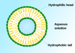
Cationic liposome
Cationic liposomes are spherical structures that contain positively charged lipids. Cationic liposomes can vary in size between 40 nm and 500 nm, and they can either have one lipid bilayer (monolamellar) or multiple lipid bilayers (multilamellar).[1] The positive charge of the phospholipids allows cationic liposomes to form complexes with negatively charged nucleic acids (DNA, mRNA, and siRNA) through ionic interactions. Upon interacting with nucleic acids, cationic liposomes form clusters of aggregated vesicles.[2] These interactions allow cationic liposomes to condense and encapsulate various therapeutic and diagnostic agents in their aqueous compartment or in their lipid bilayer.[3][4] These cationic liposome-nucleic acid complexes are also referred to as lipoplexes. Due to the overall positive charge of cationic liposomes, they interact with negatively charged cell membranes more readily than classic liposomes.[3] This positive charge can also create some issues in vivo, such as binding to plasma proteins in the bloodstream, which leads to opsonization.[5] These issues can be reduced by optimizing the physical and chemical properties of cationic liposomes through their lipid composition.[5] Cationic liposomes are increasingly being researched for use as delivery vectors in gene therapy due to their capability to efficiently transfect cells.[3][4] A common application for cationic liposomes is cancer drug delivery.[3]

History
In the 1960s, Alec D. Bangham discovered liposomes as concentric lipid bilayers surrounding an aqueous center, based on his research at the University of Cambridge Babraham Institute.[6][7] The first formulations were designed using all natural lipids.[6] In 1987, Philip Felgner published the first approach to using cationic lipids to transfect DNA into cells,[8] based on his research into cationic lipids at Syntex from 1982 to 1988.[9] Felgner introduced the first cationic lipid used for gene delivery, N-[1-(2,3-dioleyloxy)propyl]-N,N,N-trimethylammonium chloride (DOTMA).[10]
Composition
The use of cationic lipids helps to enhance the overall stability and efficiency of liposomes. Although cationic lipids themselves are able to encapsulate nucleic acids into liposomes, the transfection efficiency is low due to a process known as endosomal escape. Lipids that are capable of destabilizing endosomal membranes and facilitating endosomal escape are known as fusogenic lipids. The addition of helper lipids to the cationic lipids demonstrates a much higher transfection efficiency.[3]
Cationic liposomes with a higher transfection efficiency are composed of a cationic phospholipid, and a few neutral helper lipids. A commonly used cationic phospholipid is DOTMA, and a commonly used fusogenic helper lipid is dioleoyl phosphatidylethanolamine (DOPE). A couple of commonly used neutral helper lipids are cholesterol and polyethylene glycol (PEG)-lipid.[3] These components are all biocompatible and biodegradable in the human body, making cationic liposomes a useful gene delivery vector.[6]
Because the phospholipids each have a hydrophobic tail and a hydrophilic head group, they are able to form a lipid bilayer with the hydrophilic heads facing outwards and the hydrophobic tails facing inwards. Adding DOPE in addition to DOTMA promotes the endosomal escape of nucleic acids into the cytosol when the cationic lipid is fused to an endosome membrane.[3] The addition of cholesterol helps to stabilize the liposome and more efficiently encapsulate nucleic acids. Regulating the structure and flexibility of the lipid bilayer with cholesterol allows for a more dense assembly of phospholipids.[5] The PEG-lipid binds to the outer surface of the liposome, acting as a protective layer and reducing the formation of a protein corona.[3] The presence of PEG on the surface of the liposome drastically increases the blood circulation time of cationic liposomes.[5][6]
Manufacturing
Cationic liposomes are manufactured similarly to liposomes. There are multiple processes that can be used to form cationic liposomes, such as sonication, extrusion, and vortexing. However, the shear forces associated with these methods are capable of damaging the nucleic acids prior to encapsulation. Microfluidics is a field that is currently being looked into for the purpose of forming cationic liposomes without the shear forces and damage associated with current methods.[11]
Delivery Mechanism in vivo

Cationic liposomes can deliver nucleic acids into the cell through an endocytotic pathway or through cell membrane fusion.[3][12] Fusogenic cationic liposomes almost exclusively deliver nucleic acids through cell membrane fusion.[3] The fusion between positively charged cationic liposomes and negatively charged cell surfaces efficiently delivers the DNA directly across the plasma membrane. This process bypasses the endosomal-lysosomal route, which leads to the degradation of anionic liposome formulations.[13] Cationic liposomes in the lamellar phase deliver nucleic acids through endocytosis, specifically clathrin-mediated endocytosis (CME), caveolae-mediated endocytosis (CavME), and macropinocytosis.[3]
After administration in vivo, cationic liposomes are biodegradable due to the presence of endogenous enzymes that can digest the lipids.[14]
Applications
EndoTAG-1
Paclitaxel (PTX) is a chemotherapy medication used to treat many types of cancer, such as ovarian cancer, breast cancer, and pancreatic cancer.[7] PTX works by inhibiting the growth of tumor endothelial cells, however it has in vivo delivery issues that are caused by its unfavorable pharmacokinetic and physical properties. Studies have shown that some patients who take PTX experience adverse reactions, such as nephrotoxicity and neurotoxicity.[7][15]
SynCore Bio's EndoTAG-1 is a cationic liposome formulation with embedded PTX. The cationic lipid used in this formulation is dioleoyloxypropyltrimethylammonium (DOTAP).[16] The PTX-embedded cationic liposome interacts with the negatively charged tumor endothelial cells required for tumor angiogenesis, in order to reduce their tumor blood supply.[7][16] Through this mechanism, EndoTAG-1 is able to prevent angiogenesis in the tumor, which in turn inhibits tumor growth.[16] EndoTAG-1 is currently under phase III clinical trial investigation, and is specifically targeting adenocarcinoma of the pancreas when used in combination with gemcitabine.[17]
Issues in vivo
Cytotoxicity
The positive charge of cationic lipids has been shown to have cytotoxic effects, as these lipids can activate several pro-apoptotic and pro-inflammatory cell signaling pathways.[10] The cationic nature of these lipids is associated with the structure of the head group.[10] In particular, it was found that the quaternary ammonium head groups of some cationic lipids (e.g., DOTMA) are more cytotoxic than the peptide head groups of other cationic lipids.[10] This cytotoxic effect can be reduced by the addition of PEG on the surface of the cationic liposome.[3][5][6]
Opsonization
If delivered through intravenous administration, cationic liposomes can result in opsonization, which is an immune response that occurs when opsonins tag foreign pathogens to be eliminated through phagocytosis.[3][18] Due to their positive charge, cationic liposomes bind to various plasma proteins, forming a protein corona on their surface and completely changing their biological identity.[3] This new biological identity then causes opsonins to tag them as pathogens and encourages clearance through phagocytic clearance.[3]
See also
References
- Shah S, Dhawan V, Holm R, Nagarsenker MS, Perrie Y (2020-01-01). "Liposomes: Advancements and innovation in the manufacturing process". Advanced Drug Delivery Reviews. Advanced Liposome Research. 154–155: 102–122. doi:10.1016/j.addr.2020.07.002. PMID 32650041. S2CID 220484802.
- Elsana H, Olusanya TO, Carr-Wilkinson J, Darby S, Faheem A, Elkordy AA (October 2019). "Evaluation of novel cationic gene based liposomes with cyclodextrin prepared by thin film hydration and microfluidic systems". Scientific Reports. 9 (1): 15120. Bibcode:2019NatSR...915120E. doi:10.1038/s41598-019-51065-4. PMC 6805922. PMID 31641141. S2CID 204836762.
- Liu C, Zhang L, Zhu W, Guo R, Sun H, Chen X, Deng N (September 2020). "Barriers and Strategies of Cationic Liposomes for Cancer Gene Therapy". Molecular Therapy: Methods & Clinical Development. 18: 751–764. doi:10.1016/j.omtm.2020.07.015. PMC 7452052. PMID 32913882.
- Do HD, Ménager C, Michel A, Seguin J, Korichi T, Dhotel H, et al. (September 2020). "Development of Theranostic Cationic Liposomes Designed for Image-Guided Delivery of Nucleic Acid". Pharmaceutics. 12 (9): 854. doi:10.3390/pharmaceutics12090854. PMC 7559777. PMID 32911863.
- Inglut CT, Sorrin AJ, Kuruppu T, Vig S, Cicalo J, Ahmad H, Huang HC (January 2020). "Immunological and Toxicological Considerations for the Design of Liposomes". Nanomaterials. 10 (2): 190. doi:10.3390/nano10020190. PMC 7074910. PMID 31978968.
- Bozzuto G, Molinari A (2015-02-02). "Liposomes as nanomedical devices". International Journal of Nanomedicine. 10: 975–999. doi:10.2147/IJN.S68861. PMC 4324542. PMID 25678787.
- Beltrán-Gracia E, López-Camacho A, Higuera-Ciapara I, Velázquez-Fernández JB, Vallejo-Cardona AA (2019-12-19). "Nanomedicine review: clinical developments in liposomal applications". Cancer Nanotechnology. 10 (1): 11. doi:10.1186/s12645-019-0055-y. ISSN 1868-6966. S2CID 209452417.
- Byk G (2002). "Cationic lipid-based gene delivery". In Mahato RI, Kim SW (eds.). Pharmaceutical Perspectives of Nucleic Acid-Based Therapeutics. London: Taylor & Francis. pp. 273–303. ISBN 9780203300961.
- Jones M (22 July 1997). "Phil Felgner Interview – July 22, 1997". UC San Diego Library: San Diego Technology Archive. Regents of the University of California.
- Cui S, Wang Y, Gong Y, Lin X, Zhao Y, Zhi D, et al. (May 2018). "Correlation of the cytotoxic effects of cationic lipids with their headgroups". Toxicology Research. 7 (3): 473–479. doi:10.1039/c8tx00005k. PMC 6062336. PMID 30090597.
- Schmidt ST, Christensen D, Perrie Y (December 2020). "Applying Microfluidics for the Production of the Cationic Liposome-Based Vaccine Adjuvant CAF09b". Pharmaceutics. 12 (12): 1237. doi:10.3390/pharmaceutics12121237. PMC 7767004. PMID 33352684.
- Zelphati O, Szoka FC (October 1996). "Mechanism of oligonucleotide release from cationic liposomes". Proceedings of the National Academy of Sciences of the United States of America. 93 (21): 11493–11498. Bibcode:1996PNAS...9311493Z. doi:10.1073/pnas.93.21.11493. PMC 38085. PMID 8876163.
- "Liposomal Gene Delivery". GeneDelivery.uk. Gene Delivery. Retrieved August 23, 2017.
- Shim G, Kim MG, Park JY, Oh YK (2013-04-01). "Application of cationic liposomes for delivery of nucleic acids". Asian Journal of Pharmaceutical Sciences. Special issue on Liposomes. 8 (2): 72–80. doi:10.1016/j.ajps.2013.07.009. ISSN 1818-0876.
- Singh S, Dash AK (2009). "Paclitaxel in cancer treatment: perspectives and prospects of its delivery challenges". Critical Reviews in Therapeutic Drug Carrier Systems. 26 (4): 333–372. doi:10.1615/critrevtherdrugcarriersyst.v26.i4.10. PMID 20001890.
- Bulbake U, Doppalapudi S, Kommineni N, Khan W (March 2017). "Liposomal Formulations in Clinical Use: An Updated Review". Pharmaceutics. 9 (2): 12. doi:10.3390/pharmaceutics9020012. PMC 5489929. PMID 28346375.
- SynCore Biotechnology Co., Ltd. (2021-09-27). "A Randomized Controlled, Open Label, Adaptive Phase-3 Trial to Evaluate Safety and Efficacy of EndoTAG-1 Plus Gemcitabine Versus Gemcitabine Alone in Patients With Measurable Locally Advanced and/or Metastatic Adenocarcinoma of the Pancreas Failed on FOLFIRINOX Treatment".
{{cite journal}}: Cite journal requires|journal=(help) - Thau L, Asuka E, Mahajan K (2021). "Physiology, Opsonization". StatPearls. Treasure Island (FL): StatPearls Publishing. PMID 30480954. Retrieved 2021-11-20.