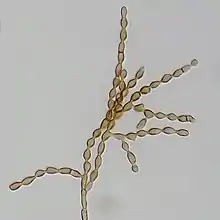
Cladosporium cladosporioides
Cladosporium cladosporioides is a darkly pigmented mold that occurs world-wide on a wide range of materials both outdoors and indoors. It is one of the most common fungi in outdoor air where its spores are important in seasonal allergic disease. While this species rarely causes invasive disease in animals, it is an important agent of plant disease, attacking both the leaves and fruits of many plants. This species produces asexual spores in delicate, branched chains that break apart readily and drift in the air. It is able to grow under low water conditions and at very low temperatures.
| Cladosporium cladosporioides | |
|---|---|
 | |
| Scientific classification | |
| Domain: | Eukaryota |
| Kingdom: | Fungi |
| Division: | Ascomycota |
| Class: | Dothideomycetes |
| Order: | Capnodiales |
| Family: | Davidiellaceae |
| Genus: | Cladosporium |
| Species: | C. cladosporioides |
| Binomial name | |
| Cladosporium cladosporioides (Fresen.) G.A.de Vries (1952) | |
| Synonyms | |
History and classification
Georg Fresenius first described Cladosporium cladosporioides in 1850, classifying it in the genus Penicillium as Penicillium cladosporioides.[1][2] In 1880 Pier Andrea Saccardo renamed the species, Hormodendrum cladosporioides.[1][2] Other early names for this taxon included Cladosporium hypophyllum, Monilia humicola and Cladosporium pisicola.[1][2] In 1952 that Gerardus Albertus de Vries transferred the species to the genus Cladosporium where it remains today.[1][2]
Growth and morphology
Cladosporium cladosporioides reproduces asexually and because no teleomorph has been identified, it is considered an exclusively anamorphic species.[3] Colonies are olive-green to olive-brown and appear velvety or powdery.[4] On a potato dextrose agar (PDA) medium, colonies are olive-grey to dull green, velvety and tufted.[5] The edges of the colony can be olive-grey to white, and feathery.[5] The colonies are diffuse and the mycelia form mats and rarely grow upwards from the surface of the colony.[5] On a malt extract agar (MEA) medium, colonies are olive-grey to olive or whitish due to the mycelia growing upwards, and seem velvety to tufted with olive-black or olive-brown edges. The mycelia can be diffuse to tufted and sometimes covers the whole colony.[5] The mycelia appear felt-like, grows flat, and can be effused and furrowed.[5] On oatmeal agar (OA) medium, colonies are olive-grey and there can be a gradient toward the edges of the colony from olive green to dull green, then olive-grey.[5] The upward growth of mycelia can be sparse to abundant and tufted.[5] The mycelia and can be loose to dense and tends to grow flat.[5] Cladosporium cladosporioides has sparse, unbranched or rarely branched, darkly-pigmented hyphae that are typically not constricted at the septa.[5] Mature conidiophores are treelike and comprise many long, branched chains of conidia.[6][7] Cladosporium cladosporioides produces brown to olive-brown coloured, solitary conidiophores that branch irregularly, forming many ramifications.[6][7] Each branch tends to be between 40–300 µm in length (exceptionally up to 350 µm) and 2–6 µm in width.[5][6] The conidiophores are thin-walled and cylindrical and are formed at the end of ascending hyphae.[8] The conidia are small, single-celled, lemon-shaped and smooth-walled.[4][6] They form long, fragile chains up to 10 conidia in length with distinctive darkened connective tissue between each spore.[4][6]
Physiology
Cladosporium cladosporioides produces antifungal metabolites targeted toward plant pathogens.[9] Three different compounds isolated from C. cladosporioides (cladosporin, isocladosporin and 5′-hydroxyasperentin), as well as a compound (5′,6-diacetyl cladosporin) synthesized from 5′-hydroxyasperentin have antifungal properties. As these compounds are effective against different types of fungi, C. cladosporioides is an important species for potential treatment and control of various plant-infecting fungi.[9] The inoculation of Venturia inaequalis, a fungus that causes apple scab on apple tree leaves, with C. cladosporioides led to decreased conidial production in V. inaequalis.[10] As this effect is seen both on younger and older leaves C. cladosporioides is effective in preventing and controlling infections of V. inaequalis in apple trees.[10] This species also produces calphostins A, B, C, D and I, which are protein kinase C inhibitors.[11][12] These calphostins have cytotoxic activity due to their ability to inhibit protein kinase C activity.[11][12]
Ecology
Cladosporium cladosporioides is a common saprotroph occurring as a secondary infection on decaying, or necrotic, parts of plants.[5] This fungus is xerophilic – growing well in low water activity environments (e.g., aW = 0.86–0.88).[13] This species is also psychrophilic, it can grow at temperatures between −10 and −3 °C (14 and 27 °F).[14] Cladosporium cladosporioides occurs outdoor environments year-round with peak spore concentration in the air occurring in summer where levels can range from 2,000 spores up to 50,000 spores per cubic meter of air.[15] It is among the most common of all outdoor airborne fungi,[15] colonizing plant materials and soil.[13] It has been found in a number of crops, such as wheat,[5] grapes,[16] strawberries,[17] peas[5] and spinach.[5] This species also grows in indoor environments,[13] where it is often associated with the growth of fungi including species of Penicillium, Aspergillus versicolor and Wallemia sebi.[15] Cladosporium cladosporioides grows well on wet building materials, paint, wallpaper and textiles,[15] as well as on paper, pulp, frescos, tiles, wet window sills and other indoor substrates[14] including salty and sugary foods.[13] Due to its tolerance of lower temperatures, C. cladosporioides can grow on refrigerated foods and colonize moist surfaces in refrigerators.[14]
Role in disease
Plants
Cladosporium cladosporioides and C. herbarum cause Cladosporium rot of red wine grapevines.[16][18] The incidence of infection is much higher when the harvest of the grapes is delayed. Over 50% of grape clusters can be affected at harvest, which greatly reduced the yield and affects the wine quality.[18][19] This delay is required in order for the phenolic compounds in the grapes to ripen and contribute to the aroma and flavour development in wine of optimum quality.[18] Symptoms of Cladosporium rot are typically observed on mature grapes and are characterized by dehydration, a small area of decay that is firm, and a layer of olive-green mould.[19] Although leaf removal reduces the incidence of infection by many species of fungi,[20] it leads to an increase in C. cladosporioides populations on grape clusters and an increase in rotten grapes at harvest.[16] Removal of diseased leaves is therefore counter-indicated in the control of this fungus.[16] The only recommendation made to avoid severe Cladosporium infections of grape clusters is to limit periods of continuous exposure to sunlight.[16]
This species has also been involved in the rotting of strawberry blossoms.[17][21][22] Infection of strawberry blossoms by C. cladosporioides has been associated with simultaneous infections by Xanthomonas fragariae (in California),[17][22] and more recently C. tenuissimum (in Korea).[21] C. cladosporioides infects the anthers, sepals, petals and pistils of the strawberry blossom[17][22] and is typically observed on older flowers with dehisced anthers and signs of senescence.[23] From 1997-2000, there was a higher proportion of misshapen fruits due to C. cladosporioides infection, and their culling affected the strawberry industry in California.[23] Infection leads to necrosis of the entire flower, or parts of it, as well as to the production of small and misshapen fruits and green-grey sporulation on the stigma.[17][21][22] A higher occurrence of infection is observed in strawberry plants cultivated outdoors than cultivated in a greenhouse.[21]
Animals
Cladosporium cladosporioides rarely causes infections in humans, although superficial infections have been reported.[4][24] It can occasionally cause pulmonary[25] and cutaneous[26] phaeohyphomycosis[4][27] and it has been isolated from cerebrospinal fluid in an immunocompromised patient.[24] This species can trigger asthmatic reactions due to the presence of allergens and beta-glucans on its spore surface.[28] In mice, living C. cladosporioides spores have induced hyperresponsiveness of the lungs, as well as an increase in eosinophils, which are white blood cells typically associated with asthmatic and allergic reactions.[28] Cladosporium cladosporioides can also induce respiratory inflammation due to the up-regulation of macrophage inflammatory protein (MIP)-2 and keratinocyte chemoattractant (KC), which are cytokines involved in the mediation of inflammation.[29] A case of mycotic encephalitis and nephritis due to C. cladosporioides has been described in a dog, resulting in altered behaviour, depression, abnormal reflexes in all 4 limbs and loss of vision.[30] Post-mortem examination indicated posterior brainstem and cerebellar lesions, confirming the causative involvement of the agent.[30]
References
- RBG Kew. "Cladosporium cladosporioides". Species Fungorum. CABI.
- International Mycological Association. "Hormodendrum cladosporioides". MycoBank Database. International Mycological Association.
- Kirk, P.M.; Cannon, Paul F.; Minter, David W.; Stalpers, J. A. (2011). Dictionary of the fungi (10th ed.). Wallingford: CABI Pub. / CSIRO. ISBN 978-1845939335.
- de Hoog, Gerrit S. (2000). Atlas of clinical fungi (2. ed.). Netherlands: Amer Society for Microbiology. pp. 1–1126. ISBN 9070351439.
- Bensch, K.; Groenewald, J.Z.; Dijksterhuis, J.; Starink-Willemse, M.; Andersen, B.; Summerell, B.A.; Shin, H.-D.; Dugan, F.M.; Schroers, H.-J.; Braun, U.; Crous, P.W. (2010). "Species and ecological diversity within the Cladosporium cladosporioides complex (Davidiellaceae, Capnodiales)". Studies in Mycology. 67: 1–94. doi:10.3114/sim.2010.67.01. PMC 2945380. PMID 20877444.
- Barron, George L. (1968). The genera of Hyphomycetes from soil. Baltimore, MD: Williams & Wilkins. ISBN 9780882750040.
- Campbell, Colin K; Johnson, Elizabeth; Warnock, David W. (2013). Identification of pathogenic fungi (2. ed.). Chichester, West Sussex: Wiley-Blackwell. pp. 100–109. ISBN 978-1118520048.
- Bensch, K.; Braun, U.; Groenewald, J.Z.; Crous, P.W. (June 2012). "The genus Cladosporium". Studies in Mycology. 72 (1): 1–401. doi:10.3114/sim0003. PMC 3390897. PMID 22815589.
- Wang, Xiaoning; Radwan, Mohamed M.; Taráwneh, Amer H.; Gao, Jiangtao; Wedge, David E.; Rosa, Luiz H.; Cutler, Horace G.; Cutler, Stephen J. (15 May 2013). "Antifungal Activity against Plant Pathogens of Metabolites from the Endophytic Fungus Cladosporium cladosporioides". Journal of Agricultural and Food Chemistry. 61 (19): 4551–4555. doi:10.1021/jf400212y. PMC 3663488. PMID 23651409.
- Köhl, Jürgen J.; Molhoek, Wilma W. M. L.; Groenenboom-de Haas, Belia B. H.; Goossen-van de Geijn, Helen H. M. (2008). "Selection and orchard testing of antagonists suppressing conidial production by the apple scab pathogen Venturia inaequalis". European Journal of Plant Pathology. 123 (4): 401–414. doi:10.1007/s10658-008-9377-z. S2CID 25678149.
- Kobayashi, Eiji; Ando, Katsuhiko; Nakano, Hirofumi; Iida, Takao; Ohno, Hiroe; Morimoto, Makoto; Tamaoki, Tatsuya (1989). "Calphostins (UCN-1028), novel and specific inhibitors of protein kinase C. I. Fermentation, isolation, physico-chemical properties and biological activities". The Journal of Antibiotics. 42 (10): 1470–1474. doi:10.7164/antibiotics.42.1470. PMID 2478514.
- Iida, Takao; Kobayashi, Eiji; Yoshida, Mayumi; Sano, Hiroshi (1989). "Calphostins, novel and specific inhibitors of protein kinase C. II. Chemical structures". The Journal of Antibiotics. 42 (10): 1475–1481. doi:10.7164/antibiotics.42.1475. PMID 2478515.
- Deshmukh, S.K.; Rai, M.K. (2005). Biodiversity of fungi : their role in human life. Enfield, NH: Science Publishers. p. 460. ISBN 1578083680.
- INSPQ. "Cladosporium cladosporioides". Institut national de santé publique Québec. Gouvernement du Québec.
- Miller, edited by Brian Flannigan, Robert A. Samson, J. David (2011). Microorganisms in home and indoor work environments : diversity, health impacts, investigation and control (2nd ed.). Boca Raton, FL: CRC Press. ISBN 9781420093346.
{{cite book}}:|first1=has generic name (help)CS1 maint: multiple names: authors list (link) - Latorre, B.A.; Briceño, E.X.; Torres, R. (January 2011). "Increase in Cladosporium spp. populations and rot of wine grapes associated with leaf removal". Crop Protection. 30 (1): 52–56. doi:10.1016/j.cropro.2010.08.022.
- Gubler, W. D.; Feliciano, A. J.; Bordas, A. C.; Civerolo, E. C.; Melvin, J. A.; Welch, N. C. (April 1999). "First Report of Blossom Blight of Strawberry Caused by Xanthomonas fragariae and Cladosporium cladosporioides in California". Plant Disease. 83 (4): 400. doi:10.1094/PDIS.1999.83.4.400A. PMID 30845607.
- Briceño, Erika X.; Latorre, Bernardo A. (December 2008). "Characterization of Cladosporium Rot in Grapevines, a Problem of Growing Importance in Chile". Plant Disease. 92 (12): 1635–1642. doi:10.1094/PDIS-92-12-1635. PMID 30764298.
- Briceño, E. X.; Latorre, B. A. (2007). "Outbreaks of Cladosporium Rot Associated with Delayed Harvest Wine Grapes in Chile". Plant Disease. 91 (8): 1060. doi:10.1094/PDIS-91-8-1060C. PMID 30780470.
- Duncan, R. A.; Stapleton, J. J.; LEAVITT, G. M. (1995). "Population dynamics of epiphytic mycoflora and occurrence of bunch rots of wine grapes as influenced by leaf removal". Plant Pathology. 44 (6): 956–965. doi:10.1111/j.1365-3059.1995.tb02653.x.
- Nam, Myeong Hyeon; Park, Myung Soo; Kim, Hyun Sook; Kim, Tae Il; Kim, Hong Gi (2015). "Cladosporium cladosporioides and C. tenuissimum Cause Blossom Blight in Strawberry in Korea". Mycobiology. 43 (3): 354–9. doi:10.5941/MYCO.2015.43.3.354. PMC 4630446. PMID 26539056.
- Gubler, W. Douglas; Feliciano, Connie J.; Bordas, Adria C.; Civerolo, Ed L.; Melvin, Jason A.; Welch, Norman C. (July 1999). "X. fragariae and C. cladosporioides cause strawberry blossom blight". California Agriculture. 53 (4): 26–28. doi:10.3733/ca.v053n04p26.
- Koike, Steven T.; Vilchez, Miguel S.; Paulus, Albert O. (2003). "Fungal Ecology of Strawberry Flower Anthers and the Saprobic Role of Cladosporium cladosporioides in Relation to Fruit Deformity Problems". HortScience. 38 (2): 246–250. doi:10.21273/HORTSCI.38.2.246.
- Kantarcioǧlu, A. S.; Yücel, A.; Hoog, G. S. (December 2002). "Case report. Isolation of Cladosporium cladosporioides from cerebrospinal fluid". Mycoses. 45 (11–12): 500–503. doi:10.1046/j.1439-0507.2002.00811.x. PMID 12472729. S2CID 44812374.
- Castro, A. S.; Oliveira, A.; Lopes, V. (31 May 2013). "Pulmonary phaeohyphomycosis: a challenge to the clinician". European Respiratory Review. 22 (128): 187–188. doi:10.1183/09059180.00007512. PMC 9487375. PMID 23728874.
- Annessi, G.; Cimitan, A.; Zambruno, G.; Silverio, A. Di (24 April 1992). "Cutaneous phaeohyphomycosis due to Cladosporium cladosporioides". Mycoses. 35 (9–10): 243–246. doi:10.1111/j.1439-0507.1992.tb00855.x. PMID 1291876. S2CID 5624047.
- Matsumoto, T.; Ajello, L.; Matsuda, T.; Szaniszlo, P.J.; Walsh, T.J. (1994). "Developments in hyalohyphomycosis and phaeohyphomycosis". Medical Mycology. 32 (s1): 329–349. doi:10.1080/02681219480000951. PMID 7722796.
- Mintz-Cole, Rachael A.; Brandt, Eric B.; Bass, Stacey A.; Gibson, Aaron M.; Reponen, Tiina; Khurana Hershey, Gurjit K. (July 2013). "Surface availability of beta-glucans is critical determinant of host immune response to Cladosporium cladosporioides". Journal of Allergy and Clinical Immunology. 132 (1): 159–169.e2. doi:10.1016/j.jaci.2013.01.003. PMC 6145803. PMID 23403046.
- Shahan, Tracy A.; Sorenson, W. G.; Paulauskis, Joseph D.; Morey, Roger; Lewis, Daniel M. (March 1998). "Concentration- and Time-dependent Upregulation and Release of the Cytokines MIP-2, KC, TNF, and MIP-1 α in Rat Alveolar Macrophages by Fungal Spores Implicated in Airway Inflammation". American Journal of Respiratory Cell and Molecular Biology. 18 (3): 435–440. doi:10.1165/ajrcmb.18.3.2856. PMID 9490662.
- Poutahidis, T.; Angelopoulou, K.; Karamanavi, E.; Polizopoulou, Z.S.; Doulberis, M.; Latsari, M.; Kaldrymidou, E. (January 2009). "Mycotic Encephalitis and Nephritis in a Dog due to Infection with Cladosporium cladosporioides". Journal of Comparative Pathology. 140 (1): 59–63. doi:10.1016/j.jcpa.2008.09.002. PMID 19064269.