DsbA
DsbA is a bacterial thiol disulfide oxidoreductase (TDOR). DsbA is a key component of the Dsb (disulfide bond) family of enzymes. DsbA catalyzes intrachain disulfide bond formation as peptides emerge into the cell's periplasm.[2]
| DsbA | |||||||
|---|---|---|---|---|---|---|---|
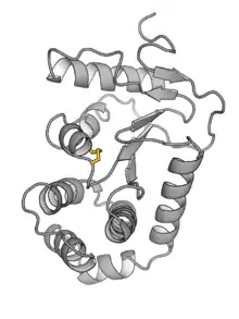
 Crystal structure of E. coli DsbA.[1] | |||||||
| Identifiers | |||||||
| Symbol | DsbA | ||||||
| NCBI gene | 948353 | ||||||
| PDB | 1A2M | ||||||
| UniProt | P0AEG4 | ||||||
| |||||||
| DSBA oxidoreductase | |||||||||||
|---|---|---|---|---|---|---|---|---|---|---|---|
| Identifiers | |||||||||||
| Symbol | DSBA | ||||||||||
| Pfam | PF01323 | ||||||||||
| InterPro | IPR001853 | ||||||||||
| |||||||||||
Structurally, DsbA contains a thioredoxin domain with an inserted helical domain of unknown function.[3] Like other thioredoxin-based enzymes, DsbA's catalytic site is a CXXC motif (CPHC in E. coli DsbA). The pair of cysteines may be oxidized (forming an internal disulfide) or reduced (as free thiols), and thus allows for oxidoreductase activity by serving as an electron pair donor or acceptor, depending on oxidation state. This reaction generally proceeds through a mixed-disulfide intermediate, in which a cysteine from the enzyme forms a bond to a cysteine on the substrate. DsbA is responsible for introducing disulfide bonds into nascent proteins. In equivalent terms, it catalyzes the oxidation of a pair of cysteine residues on the substrate protein. Most of the substrates for DsbA are eventually secreted, and include important toxins, virulence factors, adhesion machinery, and motility structures[4] DsbA is localized in the periplasm, and is more common in Gram-negative bacteria than in Gram-positive bacteria. Within the thioredoxin family, DsbA is the most strongly oxidizing member. Using glutathione oxidation as a metric, DsbA is ten times more oxidizing than protein disulfide-isomerase (the eukaryotic equivalent of DsbA). The extremely oxidizing nature of DsbA is due to an increase in stability upon reduction of DsbA, thereby imparting a decrease in energy of the enzyme when it oxidizes substrate.[5] This feature is incredibly rare among proteins, as nearly all proteins are stabilized by the formation of disulfide bonds. DsbA's highly oxidizing nature is a result of hydrogen bond, electrostatic and helix-dipole interactions that favour the thiolate over the disulfide at the active site.
After donating its disulfide bond, DsbA is regenerated by the membrane-bound protein DsbB.
References
- Guddat, LW (1998). "RCSB Protein Data Bank - RCSB PDB - 1A2M Structure Summary". Structure. 6: 757–767. doi:10.2210/pdb1a2m/pdb. Retrieved 11 July 2012.
- Kadokura H, Beckwith J (September 2009). "Detecting folding intermediates of a protein as it passes through the bacterial translocation channel". Cell. 138 (6): 1164–73. doi:10.1016/j.cell.2009.07.030. PMC 2750780. PMID 19766568.
- Guddat LW, Bardwell JC, Martin JL (June 1998). "Crystal structures of reduced and oxidized DsbA: investigation of domain motion and thiolate stabilization". Structure. 6 (6): 757–67. doi:10.1016/S0969-2126(98)00077-X. PMID 9655827.
- Heras B, Shouldice SR, Totsika M, Scanlon MJ, Schembri MA, Martin JL (March 2009). "DSB proteins and bacterial pathogenicity". Nature Reviews. Microbiology. 7 (3): 215–25. doi:10.1038/nrmicro2087. PMID 19198617. S2CID 34433130.
- Zapun A, Bardwell JC, Creighton TE (May 1993). "The reactive and destabilizing disulfide bond of DsbA, a protein required for protein disulfide bond formation in vivo". Biochemistry. 32 (19): 5083–92. CiteSeerX 10.1.1.455.5367. doi:10.1021/bi00070a016. PMID 8494885.