Ethmoid sinus
The ethmoid sinuses or ethmoid air cells of the ethmoid bone are one of the four paired paranasal sinuses.[1] Unlike the other three pairs of paranasal sinuses which consist of one or two large cavities, the ethmoidal sinuses entail a number of small air-filled cavities ("air cells").[2] The cells are located within the lateral mass (labyrinth) of each ethmoid bone and are variable in both size and number.[1] The cells are grouped into anterior, middle, and posterior groups; the groups differ in their drainage modalities,[2] though all ultimately drain into either the superior or the middle nasal meatus[3] of the lateral wall of the nasal cavity.
| Ethmoid sinus | |
|---|---|
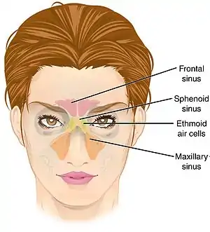 Frontal view of paranasal sinuses | |
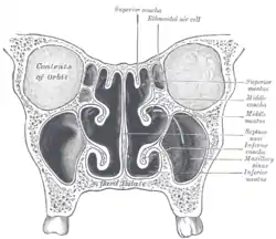 Coronal section of nasal cavities. | |
| Details | |
| Nerve | posterior ethmoidal nerve |
| Identifiers | |
| Latin | Cellulae ethmoidales, labyrinthi ethmoidales |
| MeSH | D005005 |
| TA98 | A06.1.03.005 |
| TA2 | 728, 3180 |
| FMA | 84115 |
| Anatomical terms of bone | |
Structure
The ethmoid air cells consist of numerous thin-walled cavities in the ethmoidal labyrinth[4] that represent invaginations of the mucous membrane of the nasal wall into the ethmoid bone.[3] They are situated between the superior parts of the nasal cavities and the orbits, and are separated from these cavities by thin bony lamellae.[4]
There are 5-15 air cells in either ethmoid bone in the adult, with a combined volume of 2-3mL.[5]
Drainage
- The anterior ethmoidal cells drain (directly or indirectly[3]) into the middle nasal meatus by way of the ethmoidal infundibulum.[4][3][5]
- The middle ethmoidal cells drain directly into the middle nasal meatus.[3]
- The posterior ethmoidal cells drain directly into the superior nasal meatus[3][5] at the sphenoethmoidal recess;[5] sometimes, one or more opens into the sphenoidal sinus.[4]
Haller cells
Haller cells are infraorbital ethmoidal air cells lateral to the lamina papyracea. These may arise from the anterior or posterior ethmoidal sinuses.
Lamellae
The ethmoidal labyrinth is divided by multiple obliquely oriented, parallel lamellae. The first lamellae is equivalent to the uncinate process of ethmoid bone, the second corresponds the ethmoid bulla, and the third is the basal lamella, and the fourth is equivalent to the superior nasal concha.[5]
The anterior and posterior ethmoid cells are separated by the basal lamella[6][5] (also known as the ground lamella).[5] It is one of the bony divisions of the ethmoid bone and is mostly contained inside the ethmoid labyrinth. The basal lamella is continuous medially with the bony middle nasal concha.[6] Anteriorly, it vertically inserts into the ethmoid crest; the middle part attaches obliquely into the orbital lamina of ethmoid bone (lamina papyricea) while the posterior part attaches into the orbital lamina horizontally.[5]
Innervation
The ethmoidal air cells receive sensory innervation from the anterior and the posterior ethmoidal nerve (which are ultimately derived from the ophthalmic branch (CN V1) of the trigeminal nerve (CN V)),[3] and the orbital branches of the pterygopalatine ganglion, which carry the postganglionic parasympathetic nerve fibers for mucous secretion from the facial nerve.
Clinical significance
Acute ethmoiditis in childhood and ethmoidal carcinoma may spread superiorly causing meningitis and cerebrospinal fluid leakage or it may spread laterally into the orbit causing proptosis and diplopia.[8]
Additional images
 Ethmoid sinus. Ethmoidal air cells.Deep dissection. Superior view.
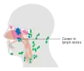 Ethmoid sinus cancer that has spread to the lymph nodes
References
![]() This article incorporates text in the public domain from page 154 of the 20th edition of Gray's Anatomy (1918)
This article incorporates text in the public domain from page 154 of the 20th edition of Gray's Anatomy (1918)
- Illustrated Anatomy of the Head and Neck, Fehrenbach and Herring, Elsevier, 2012, page 64
- Morton, David A. (2019). The Big Picture: Gross Anatomy. K. Bo Foreman, Kurt H. Albertine (2nd ed.). New York. p. 246. ISBN 978-1-259-86264-9. OCLC 1044772257.
{{cite book}}: CS1 maint: location missing publisher (link) - Moore, Keith L.; Dalley, Arthur F.; Agur, Anne M. R. (2017). Essential Clinical Anatomy (6th ed.). Lippincott Williams & Wilkins. p. 968. ISBN 978-1496347213.
- Otorhinolaryngology, Head and Neck Surgery, Anniko, Springer, 2010, page 188
- Cappello, Zachary J.; Minutello, Katrina; Dublin, Arthur B. (2023), "Anatomy, Head and Neck, Nose Paranasal Sinuses", StatPearls, Treasure Island (FL): StatPearls Publishing, PMID 29763001, retrieved 2023-07-04
- Hechl, Peter S.; Setliff, Reuben C.; Tschabitscher, Manfred (1997). "The ethmoid bone and middle turbinate". Endoscopic Anatomy of the Paranasal Sinuses. Springer Vienna. pp. 9–28. doi:10.1007/978-3-7091-6536-2_2. ISBN 978-3-7091-7345-9.
- Moore, K.L Et al(2014). Clinically Oriented Anatomy. Baltimore: Page960
- Human Anatomy, Jacobs, Elsevier, 2008, page 210
External links
- Anatomy figure: 33:04-07 at Human Anatomy Online, SUNY Downstate Medical Center
- Anatomy photo:33:st-0711 at the SUNY Downstate Medical Center