Focal plane tomography
In radiography, focal plane tomography[1] is tomography (imaging a single plane, or slice, of an object) by simultaneously moving the X-ray generator and X-ray detector so as to keep a consistent exposure of only the plane of interest during image acquisition. This was the main method of obtaining tomographs in medical imaging until the late-1970s. It has since been largely replaced by more advanced imaging techniques such as CT and MRI. It remains in use today in a few specialized applications, such as for acquiring orthopantomographs of the jaw in dental radiography.
| Focal plane tomography | |
|---|---|
 An orthopantomograph, which uses focal plane tomography. | |
| Purpose | tomography imaging a single plane/slice |
Focal plane tomography’s development began in the 1930s as a means of reducing the problem of superimposition of structures which is inherent to projectional radiography.[2] It was invented in parallel by, among others, by the French physician Bocage, the Italian radiologist Alessandro Vallebona and the Dutch radiologist Bernard George Ziedses des Plantes.[3]
Technique
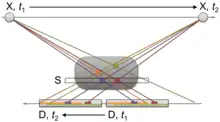
Focal plane tomography generally uses mechanical movement of an X-ray source and film in unison to generate a tomogram using the principles of projective geometry.[4] Synchronizing the movement of the radiation source and detector which are situated in the opposite direction from each other causes structures which are not in the focal plane being studied to blur out.
Limitations
The blurring provided by focal plane tomography is only marginally effective, since it only occurs in the X plane. Moreover, since focal plane tomography uses plain X-rays, it is not particularly effective at resolving soft tissues.
The increased availability and power of computers in the 1960s and 70s gave rise to new imaging techniques such as CT and MRI which use computational (in addition to or in lieu of mechanical) methods to acquire and process tomographic image data, and which do not suffer from the limitations of focal plane tomography.
Variants
Initially focal plane tomography used simple linear movements. The technique advanced through the mid-twentieth century however, steadily producing sharper images, and with a greater ability to vary the thickness of the cross-section being examined.[4] This was achieved through the introduction of more complex, pluridirectional devices that can move in more than one plane and perform more effective blurring.
Linear tomography

This is the most basic form of conventional tomography. The X-ray tube moved from point "A" to point "B" above the patient, while the detector (such as cassette holder or "bucky") moves simultaneously under the patient from point "B" to point "A".[5] The fulcrum, or pivot point, is set to the area of interest. In this manner, the points above and below the focal plane are blurred out, just as the background is blurred when panning a camera during exposure. Rarely used, and has largely been replaced by computed tomography (CT).
Poly tomography
| External video | |
|---|---|
This was achieved using a more advanced X-ray apparatus that allows for more sophisticated and continuous movements of the X-ray tube and film. With this technique, a number of complex synchronous geometrical movements could be programmed, such as hypocycloidic, circular, figure 8, and elliptical. Philips Medical Systems for example produced one such device called the 'Polytome'.[4] This pluridirectional unit was still in use into the 1990s, as its resulting images for small or difficult physiology, such as the inner ear, were still difficult to image with CTs at that time. As the resolution of CT scanners got better, this procedure was taken over by CT.[6]
Zonography
This is a variant of linear tomography, where a limited arc of movement is used, resulting in less blurring than linear tomography.[7] It is still used in some centres for visualising the kidney during an intravenous urogram (IVU),[8] though it too is being supplanted by CT.[9][10]
Panoramic radiograph
Panoramic radiography is the only common tomographic examination still in use. This makes use of a complex movement to allow the radiographic examination of the mandible, as if it were a flat bone.[11] It is commonly performed in dental practices and is often referred to as a "Panorex", though this is a trademark of a specific company and not a generic term.
See also
References
- Pickens, D. R.; Price, R. R.; Patton, J. A.; Erickson, J. J.; Rollo, F. D.; Brill, A. B. (1980). "Focal-Plane Tomography Image Reconstruction". IEEE Transactions on Nuclear Science. 27 (1): 489–492. Bibcode:1980ITNS...27..489P. doi:10.1109/TNS.1980.4330874. ISSN 0018-9499. S2CID 30852566.
- Kevles, Bettyann (1997). Naked to the Bone: Medical Imaging in the Twentieth Century. Rutgers University Press. p. 108. ISBN 9780813523583.
- Van Gijn, Jan; Gijselhart, Joost P. (2010-06-23). "Ziedses des Plantes: uitvinder van planigrafie en subtractie" (PDF). Nederlands Tijdschrift voor Geneeskunde (in Dutch).
- Littleton, J.T. "Conventional Tomography". A History of the Radiological Sciences (PDF). American Roentgen Ray Society. Retrieved 11 January 2014.
- Allisy-Roberts, Penelope; Williams, Jerry R. (2007). Farr's Physics for Medical Imaging. Elsevier Health Sciences. p. 76. ISBN 978-0702028441.
- Lane, John I.; Lindell, E. Paul; Witte, Robert J.; DeLone, David R.; Driscoll, Colin L. W. (January 2006). "Middle and Inner Ear: Improved Depiction with Multiplanar Reconstruction of Volumetric CT Data". RadioGraphics. 26 (1): 115–124. doi:10.1148/rg.261055703. PMID 16418247.
- Ettinger, Alice; Fainsinger, Maurice H. (July 1966). "Zonography in Daily Radiological Practice". Radiology. 87 (1): 82–86. doi:10.1148/87.1.82. PMID 5940479.
- Daniels, S.J.; Brennan, P.C. (May 1996). "A comparison of tomography and zonography during intravenous urography". Radiography. 2 (2): 99–109. doi:10.1016/S1078-8174(96)90002-4.
- Whitfield, Ahn; Whitfield, HN (January 2006). "Is There a Role for the Intravenous Urogram in the 21st Century?". Annals of the Royal College of Surgeons of England. 88 (1): 62–65. doi:10.1308/003588406X83168. PMC 1963625. PMID 16460641.
- Whitley, A. Stewart; Jefferson, Gail; Holmes, Ken; Sloane, Charles; Anderson, Craig; Hoadley, Graham (2015-07-28). Clark's Positioning in Radiography 13E. CRC Press. p. 526. ISBN 9781444165050.
- Ghom, Anil (2008). Textbook of Oral Radiology (1st ed.). Elsevier India. p. 460. ISBN 9788131211489.
External links
 Media related to Focal plane tomography at Wikimedia Commons