Non-ossifying fibroma
A non-ossifying fibroma (NOF) is a benign bone tumor of the osteoclastic giant cell-rich tumor type.[1] It generally occurs in the metaphysis of long bones in children and adolescents.[2] Typically, there are no symptoms unless there is a fracture.[2] It can occur as part of a syndrome such as when multiple non-ossifying fibromas occur in neurofibromatosis, or Jaffe–Campanacci syndrome in combination with cafe-au-lait spots, mental retardation, hypogonadism, eye and cardiovascular abnormalities.[2]
| Non-ossifying fibroma | |
|---|---|
| Other names | Fibroxanthoma |
 | |
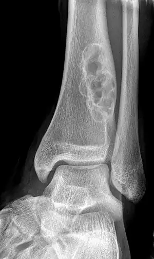
| X-ray of nonossifying fibroma of distal tibia. | |
| Specialty | Rheumatology |
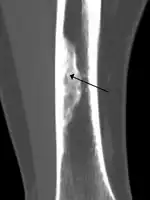
Diagnosis is by X-ray or MRI, usually when investigating a person for something else.[2] Medical imaging typically shows a well defined radiolucent lesion, with a distinct multilocular appearance, sometimes looking like bubbles.[2] It is usually around 1–2 cm in size, but be as large as 7 cm.[3] They consist of foci consist of collagen rich connective tissue, fibroblasts, histiocytes and osteoclasts.[2] Usually no treatment is required.[1] Surgical curettage and bone grafting may be required if it is large.[3]
It is found in 30–40% of children and adolescents, but rare in adults as most have resolved by this time.[2] They do not become malignant.[2] It affects twice as many males as females.[2] A NOF was identified on the mandible of Qafzeh 9, an early anatomically modern human dated to 90–100 000 yrs B.P.[4]
Signs and symptoms
Most people with non-ossifying fibroma have no symptoms.[1] If the tumor is large, there may be pain over the affected area, a pathological fracture, and the affected limb might not function properly.[1] It can occur as part of a syndrome such as when multiple non-ossifying fibromas occur in neurofibromatosis, or Jaffe–Campanacci syndrome in combination with cafe-au-lait spots, mental retardation, hypogonadism, eye and cardiovascular abnormalities.[2]
Diagnosis
It is usually diagnosed by x-ray or MRI, when investigating another problem.[1] The tumor presents as a well defined radiolucent lesion, with a distinct multilocular appearance, sometimes looking like a "soap bubble".[5] If small and no symptoms, then biopsy is not needed.[1]
Additional images

See also
References
- WHO Classification of Tumours Editorial Board, ed. (2020). "Non-ossifying fibroma". Soft Tissue and Bone Tumours: WHO Classification of Tumours. Vol. 3 (5th ed.). Lyon (France): International Agency for Research on Cancer. pp. 447–448. ISBN 978-92-832-4503-2.
- Murali, Sundaram; Ilaslan, Hakan; Holden, Darlene M. (2015). "2. An imaging approach to bone tumors". In Santini-Araujo, Eduardo; Kalil, Ricardo K.; Bertoni, Franco; Park, Yong-Koo (eds.). Tumors and Tumor-Like Lesions of Bone: For Surgical Pathologists, Orthopedic Surgeons and Radiologists. Springer. p. 15. ISBN 978-1-4471-6577-4.
- Paulos, Jaime (2021). "Non Ossifying Fibroma". Bone Tumors: Diagnosis and Therapy Today. Springer. p. 139. doi:10.1007/978-1-4471-7501-8_22. ISBN 978-1-4471-7501-8. S2CID 238034517.
- Coutinho Nogueira D, Dutour O, Coqueugniot H, Tillier A.-m., (2019) Qafzeh 9 mandible (ca 90–100 kyrs BP, Israel) revisited : μ-CT and 3D reveal new pathological conditions, International Journal of Paleopathology, Vol 26, pp.104-110, https://doi.org/10.1016/j.ijpp.2019.06.002
- Ahn, Leah; O'Donnell, Patrick. "Non-Ossifying Fibroma - Pathology - Orthobullets". www.orthobullets.com. Retrieved 3 August 2021.
External links
 Media related to Nonossifying fibroma at Wikimedia Commons
- Wheeless' Textbook of Orthopaedics: Nonossifying Fibroma