Perfusion CT
Perfusion CT or CT Perfusion is a type of Perfusion Scanning using Computed Tomography. It is helpful in evaluation of the vascularity of a tissue in the body. In this the temporal changes in the tissue density are measured which gives the information about the vascularity of the tissue. In CT perfusion injection of contrast media is given and then the scan is taken. The acquired data are then post-processed to obtain perfusion maps with different parameters, such as BV (blood volume), BF (blood flow), MTT (mean transit time) and TTP (time to peak).[1][2]
| Perfusion CT | |
|---|---|
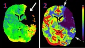
 CT perfusion with flow and volume maps in cerebral infarction | |
| Synonyms | CT Perfusion |
| Purpose | Perfusion Scanning using CT |
| Based on | Temporal changes in tissue attenuation after injection of Contrast |
Clinical use
Acute Ischemic Stroke
CT Perfusion plays an important role in the assessment of Acute Ischemia Stroke. It is used to create maps of blood flow, blood volume and mean transit time to assess the tissue and to differentiate between core and penumbra in stroke.[3]
References
- Saremi, Farhood (2015-05-22). Perfusion Imaging in Clinical Practice: A Multimodality Approach to Tissue Perfusion Analysis. Lippincott Williams & Wilkins. ISBN 978-1-4963-1804-6.
- Diagnostic radiology. Recent advances and applied physics in imaging. Arun Kumar Gupta (Second ed.). New Delhi. 2013. ISBN 978-93-5090-497-8. OCLC 872632898.
{{cite book}}: CS1 maint: location missing publisher (link) CS1 maint: others (link) - "Eckert B, Küsel T, Leppien a et al. Clinical outcome and imaging follow-up in acute stroke patients with normal perfusion CT and normal CT angiography. Neuroradiology 2011; 53: 79–88". Neuroradiologie Scan. 1 (1): 22–23. 2011-10-12. doi:10.1055/s-0030-1256919. ISSN 1616-9697. S2CID 260379976.