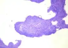
Perianal gland tumor
A perianal gland tumor is a type of tumor found near the anus in dogs that arises from specialized glandular tissue found in the perineum.[1] It is also known as a hepatoid tumor because of the similarity in cell shape to hepatocytes (liver cells). It is most commonly seen in intact (not neutered) dogs and is the third most common tumor type in intact male dogs.[2] There are two types of perianal gland tumors, perianal gland adenomas, which are benign, and perianal gland adenocarcinomas, which are malignant. Both have receptors for testosterone.[3] Perianal gland adenomas are three times more likely to be found in intact male dogs than females, and perianal gland adenocarcinomas are ten times more common in male dogs than females.[4] The most commonly affected breeds for adenomas are the Siberian Husky, Cocker Spaniel, Pekingese, and Samoyed; for adenocarcinomas the most commonly affected breeds are the Siberian Husky, Bulldog, and Alaskan Malamute.[4]

Perianal gland tumors are located most commonly in the skin around the anus, but can also be found on the tail or groin. Adenomas are more common, making up 91 percent of perianal gland tumors in one study.[5] Adenomas and adenocarcinomas look alike, both being round, pink and usually less than three centimeters in width. Adenocarcinomas are more likely to be multiple and invasive into the underlying tissue, and they can metastasize to the lymph nodes, liver, and lungs.
Both types should be removed and sent to a pathologist for identification. However, 95 percent of perianal gland adenomas will disappear after neutering the dog.[5] Rem
emoving the tumor and neutering the dog at the same time will help prevent recurrnce. Dogs with perianal gland adenocarcinomas should be treated with aggressive surgery and the radiation therapy and chemotherapy if necessary.
References
- Kirpensteijn, Jolle (Jan 2006). "Treatment of perianal and anal sac tumors" (PDF). Proceedings of the North American Veterinary Conference. Retrieved 2007-03-27.
- Petterino C, Martini M, Castagnaro M (2004). "Immunohistochemical detection of growth hormone (GH) in canine hepatoid gland tumors". J Vet Med Sci. 66 (5): 569–72. doi:10.1292/jvms.66.569. PMID 15187372.
- Pisani G, Millanta F, Lorenzi D, Vannozzi I, Poli A (2006). "Androgen receptor expression in normal, hyperplastic and neoplastic hepatoid glands in the dog". Res Vet Sci. 81 (2): 231–6. doi:10.1016/j.rvsc.2005.11.001. PMID 16427103.
- "Hepatoid Gland Tumors". The Merck Veterinary Manual. 2006. Retrieved 2007-03-27.
- Morrison, Wallace B. (1998). Cancer in Dogs and Cats (1st ed.). Williams and Wilkins. ISBN 0-683-06105-4.