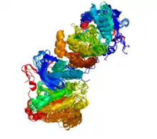
ProtCID
The Protein Common Interface Database (ProtCID) is a database of similar protein-protein interfaces in crystal structures of homologous proteins.[1][5]
| Content | |
|---|---|
| Description | Similar interactions of homologous proteins in multiple crystal forms |
| Contact | |
| Research center | Fox Chase Cancer Center |
| Laboratory | Institute for Cancer Research |
| Authors | Qifang Xu, Roland Dunbrack |
| Primary citation | Xu & Dunbrack (2011)[1] |
| Release date | 2010 |
| Access | |
| Website | http://dunbrack2.fccc.edu/protcid |

Its main goal is to identify and cluster homodimeric and heterodimeric interfaces observed in multiple crystal forms of homologous proteins. Such interfaces, especially of non-identical proteins or protein complexes, have been associated with biologically relevant interactions.[6]
A common interface in ProtCID indicates chain-chain or domain-domain interactions that occur in different crystal forms. All protein sequences of known structure in the Protein Data Bank (PDB)[7] are assigned a ”Pfam chain architecture”, which denotes the ordered Pfam[8] assignments for that sequence, e.g. (Pkinase) or (Cyclin_N)_(Cyclin_C). Homodimeric interfaces in all crystals that contain particular domain or chain architectures are compared, regardless of whether there are other protein types in the crystals. All interfaces between two different Pfam domains or Pfam architectures in all PDB entries that contain them are also compared (e.g., (Pkinase) and (Cyclin_N)_(Cyclin_C) ). For both homodimers and heterodimers, the interfaces are clustered into common interfaces based on a similarity score.
ProtCID reports the number of crystal forms that contain a common interface, the number of PDB entries, the number of PDB and PISA[9] biological assembly annotations that contain the same interface, the average surface area, and the minimum sequence identity of proteins that contain the interface. ProtCID provides an independent check on publicly available annotations of biological interactions for PDB entries.
ProtCID also contains interface clusters between protein domains and peptides, nucleic acids, and ligands.
See also
References
- Xu, Q.; Dunbrack, R. L. (2010). "The protein common interface database (ProtCID)—a comprehensive database of interactions of homologous proteins in multiple crystal forms". Nucleic Acids Research. 39 (Database issue): D761–70. doi:10.1093/nar/gkq1059. PMC 3013667. PMID 21036862.
- Zhang, X.; Gureasko, J.; Shen, K.; Cole, P. A.; Kuriyan, J. (2006). "An Allosteric Mechanism for Activation of the Kinase Domain of Epidermal Growth Factor Receptor". Cell. 125 (6): 1137–1149. doi:10.1016/j.cell.2006.05.013. PMID 16777603.
- Aertgeerts, K.; Skene, R.; Yano, J.; Sang, B. -C.; Zou, H.; Snell, G.; Jennings, A.; Iwamoto, K.; Habuka, N.; Hirokawa, A.; Ishikawa, T.; Tanaka, T.; Miki, H.; Ohta, Y.; Sogabe, S. (2011). "Structural Analysis of the Mechanism of Inhibition and Allosteric Activation of the Kinase Domain of HER2 Protein". Journal of Biological Chemistry. 286 (21): 18756–18765. doi:10.1074/jbc.M110.206193. PMC 3099692. PMID 21454582.
- Qiu, C.; Tarrant, M. K.; Choi, S. H.; Sathyamurthy, A.; Bose, R.; Banjade, S.; Pal, A.; Bornmann, W. G.; Lemmon, M. A.; Cole, P. A.; Leahy, D. J. (2008). "Mechanism of Activation and Inhibition of the HER4/ErbB4 Kinase". Structure. 16 (3): 460–467. doi:10.1016/j.str.2007.12.016. PMC 2858219. PMID 18334220.
- Xu, Q; Dunbrack, RL (5 February 2020). "ProtCID: a data resource for structural information on protein interactions". Nature Communications. 11 (1): 711. Bibcode:2020NatCo..11..711X. doi:10.1038/s41467-020-14301-4. PMC 7002494. PMID 32024829.
- Xu, Qifang; Canutescu, Adrian A.; Wang, Guoli; Shapovalov, Maxim; Obradovic, Zoran; Dunbrack, Roland L. (2008). "Statistical Analysis of Interface Similarity in Crystals of Homologous Proteins". Journal of Molecular Biology. 381 (2): 487–507. doi:10.1016/j.jmb.2008.06.002. PMC 2573399. PMID 18599072.
- Berman, H. M.; Battistuz, T.; Bhat, T. N.; Bluhm, W. F.; Bourne, P. E.; Burkhardt, K.; Feng, Z.; Gilliland, G. L.; Iype, L.; Jain, S.; Fagan, P.; Marvin, J.; Padilla, D.; Ravichandran, V.; Schneider, B.; Thanki, N.; Weissig, H.; Westbrook, J. D.; Zardecki, C. (2002). "The Protein Data Bank". Acta Crystallographica Section D. 58 (Pt 6 No 1): 899–907. doi:10.1107/S0907444902003451. PMID 12037327.
- Punta, M.; Coggill, P. C.; Eberhardt, R. Y.; Mistry, J.; Tate, J.; Boursnell, C.; Pang, N.; Forslund, K.; Ceric, G.; Clements, J.; Heger, A.; Holm, L.; Sonnhammer, E. L. L.; Eddy, S. R.; Bateman, A.; Finn, R. D. (2011). "The Pfam protein families database". Nucleic Acids Research. 40 (Database issue): D290–D301. doi:10.1093/nar/gkr1065. PMC 3245129. PMID 22127870.
- Krissinel, E.; Henrick, K. (2007). "Inference of Macromolecular Assemblies from Crystalline State". Journal of Molecular Biology. 372 (3): 774–797. doi:10.1016/j.jmb.2007.05.022. PMID 17681537.