STIL
SCL-interrupting locus protein is a protein that in humans is encoded by the STIL gene. STIL is present in many different cell types and is essential for centriole biogenesis. This gene encodes a cytoplasmic protein implicated in regulation of the mitotic spindle checkpoint, a regulatory pathway that monitors chromosome segregation during cell division to ensure the proper distribution of chromosomes to daughter cells. The protein is phosphorylated in mitosis and in response to activation of the spindle checkpoint, and disappears when cells transition to G1 phase. It interacts with a mitotic regulator, and its expression is required to efficiently activate the spindle checkpoint.
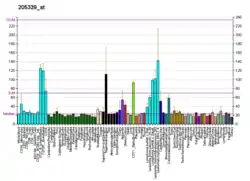
It is proposed to regulate Cdc2 kinase activity during spindle checkpoint arrest. Chromosomal deletions that fuse this gene and the adjacent locus commonly occur in T cell leukemias, and are thought to arise through illegitimate recombination events. Multiple transcript variants encoding different isoforms have been found for this gene. Multiple types of cancer produce STIL, and its expression is linked to an increased mitotic index and cancer development.[5] Hedgehog family-mediated signaling events are one of its associated pathways. The development and function of the nervous system are impacted by STIL.[6] The sequence of STIL gene is highly conserved in vertebrate species .Both fetal and adult tissues express the STIL gene. Its expression levels fluctuate with the cell cycle, making it challenging to detect in a complete tissue, particularly if the cells are not synchronized.
Gene location
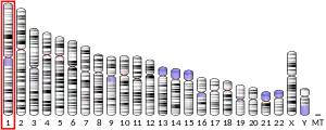
The human STIL gene is located on the (p) arm of chromosome 1. It mapped the STIL gene to chromosome 1p33 based on an alignment of the STIL sequence with the genomic sequence. STIL gene contains 20 exons, including alternatively spliced exons 13A and 13B and 18A and 18B. The coding region begins in exon 3. The human SIL gene encodes a 1287-amino acid cytosolic protein.
Functions and mechanism
Numerous cancer types are affected by STIL overexpression which has been linked to chromosomal instability. It plays a part in neural development and function. STIL plays a crucial role in cell mitosis and centriole replication. The early stages of the cell cycle see a slow increase in STIL expression, a peak in the middle, and a sharp decline in the latter stages. When cellular proliferation is inhibited by serum deprivation, contact inhibition, or the promotion of terminal differentiation, STIL is expressed in the proliferating cells and is down-regulated. STIL has been interacted with CDK1, PLK4, and SAS-6. STIL has a role in the Sonic hedgehog (Shh) pathway. STIL regulates the transcription of Shh-target gene Gli1[7] .The suppressor-of-fused homolog (SUFU) and GLI1 are examples of conserved Shh signaling elements with which the C terminus of STIL can engage. The activation of Shh-GLI1 cascades is caused by STIL's interaction with SUFU, which prevents SUFU from acting as a repressor of GLI1.
Normally, GLI1 binds to the cytoplasmic protein SUFU to form heterodimers. The transcription of the Gli1 gene is blocked because the heterodimers cannot be translocated to nucleus. The binding of SUFU by STIL during STIL expression releases GLI1 from SUFU repression. Gene transcription can then begin as GLI1 enters the nucleus. The transcription of Gli1 cannot begin if STIL is altered. Normally, STIL to bind SUFU, relieve SUFU's inhibition of GLI1, and then allow GLI1 to go to the nucleus for gene transcription. The inability of the SUFU-GLI1 heterodimers prevents the completion of Shh downstream signaling transduction when STIL is mutated.
Role of STIL in cancer
Numerous malignancies have been identified to have STIL disorders, which have fueled carcinogenesis. Copy number variation, mutation, and DNA methylation all had an impact on STIL's dysregulated expression. The expression of STIL was inversely linked with numerous ciliogenesis-related genes. The equilibrium of STIL expression is crucial for the development of primary cilia. STIL silencing might facilitate the development of primary cilia and prevent the production of cell cycle-related proteins. There are no primary cilia when STIL expression is completely lost. Increased cancer metastatic potential is linked to STIL overexpression. STIL has associated with various cancers including lung cancer, colon cancer, pancreatic cancer, prostate adenocarcinoma, and ovarian cancer. The production of mitotic spindles, as well as SHH signaling and the operation of its interactors, are all likely impacted by STIL overexpression, which is linked to a high histopathological mitotic index in tumors. Overexpression of STIL may function as oncogenes and cause cancer by encouraging spindle abnormalities. Spindle orientation control is lost due to disordered mitotic spindles caused by STIL downregulation. This may lead to a reduction in the number of cortical progenitors by cell death or premature differentiation. PLK4 overexpression also causes centrosome amplification and aneuploidy, which reduce brain volume as a result of cell death . Apoptosis inhibition in this setting results in an accumulation of aneuploid cells that are unable to proliferate effectively, causing premature neural differentiation, whereas PLK4 overexpression in environment induces skin cancer.
As a PLK4 downstream effector, STIL may possibly indirectly affect cancer. Malignancies such juvenile medulloblastoma, breast tumors, and colorectal cancer have all been linked to elevated PLK4 expression. Multiple organs develop spontaneous tumors as a result of PLK4 overexpression. It is unclear if STIL expression is necessary for this trait. PLK4 remodels the cytoskeleton and may be important for cancer invasion and metastasis because STIL binds to PLK4 in the cytoplasm. As a result, STIL expression levels may have an impact on PLK4 cytoplasmic activity. PLK4 depletion is associated with an increase in E-cadherin expression and a reduction in metastasis.
In addition to these conditions, CYCLIN B is frequently elevated in primary breast cancer, esophageal squamous cell carcinoma, laryngeal squamous cell carcinoma, and colorectal carcinoma. Downregulation of STIL inhibits tumor growth in vivo by lowering CDK1/CYCLIN B activity, delaying G2-M transition, and preventing G2-M transition. While elevating STIL might encourage CDK1/CYCLIN B activity and unintentionally contribute to CYCLIN B-dependent proliferation in tumor cells. The absence of STIL also causes an increase of Chfr and a decrease in PLK1, which activates the CDC25c phosphatase. Thus, this route may be able to regulate cell division independent of its essential function in centriole duplication.
Role of STIL in neural development
The pattern of STIL expression during the fetal stages supports the link between this gene and cell proliferation. At 15 postconceptional weeks, STIL is more strongly expressed in the ganglionic eminence, the rostral migratory stream, the ventricular and sub ventricular zones of the forebrain.[6] While it is less expressed in the intermediate zone, sub plate, cortical plate, marginal zone, and sub granular layer. The manifestation of this pattern is still present at 21 postconceptional weeks, but it is less prominent in the sub ventricular region.
Although the expression of SAS-6 and STIL differs in some areas of the cortical plate, PLK4, SAS-6, and CPAP also often exhibit this pattern of expression. The exterior granule layer and areas of the rhombic lip of the cerebellum express STIL, PLK4, and SAS-6 but not CPAP. However, none of these genes are expressed in the migratory streams of the hindbrain, the ventricular matrix zone of the cerebellum, or the transitory Purkinje cell cluster.
When the head circumference is less than the age-specific and gender-adjusted mean by more than two standard deviations (S.D.s) at birth, microcephaly (small brain size) is inferred. Primary microcephaly is the term used to describe genetic microcephalies that can be seen in pregnancy. The majority of them, known as microcephalic dwarfism, are autosomal recessive and include I solitary variants known as Microcephaly Primary Hereditary (MCPH), and (ii) types linked to growth retardation. A MCPH phenotype is linked to the majority of STIL mutations found in patients, and STIL is known as MCPH7. Both an increase and a decrease in STIL protein levels during the cell cycle have an impact on centriole control and cause microcephaly.
References
- GRCh38: Ensembl release 89: ENSG00000123473 - Ensembl, May 2017
- GRCm38: Ensembl release 89: ENSMUSG00000028718 - Ensembl, May 2017
- "Human PubMed Reference:". National Center for Biotechnology Information, U.S. National Library of Medicine.
- "Mouse PubMed Reference:". National Center for Biotechnology Information, U.S. National Library of Medicine.
- Patwardhan, Dhruti; Mani, Shyamala; Passemard, Sandrine; Gressens, Pierre; El Ghouzzi, Vincent (2018-01-19). "STIL balancing primary microcephaly and cancer". Cell Death & Disease. 9 (2): 65. doi:10.1038/s41419-017-0101-9. ISSN 2041-4889. PMC 5833631. PMID 29352115.
- Li, Lei; Liu, Congcong; Carr, Aprell L. (2019-04-25). "STIL: a multi-function protein required for dopaminergic neural proliferation, protection, and regeneration". Cell Death Discovery. 5 (1): 90. doi:10.1038/s41420-019-0172-8. ISSN 2058-7716. PMC 6484007. PMID 31044090.
- Kasai, Kenji; Inaguma, Shingo; Yoneyama, Akiko; Yoshikawa, Kazuhiro; Ikeda, Hiroshi (2008-10-01). "SCL/TAL1 Interrupting Locus Derepresses GLI1 from the Negative Control of Suppressor-of-Fused in Pancreatic Cancer Cell". Cancer Research. 68 (19): 7723–7729. doi:10.1158/0008-5472.CAN-07-6661. ISSN 0008-5472. PMID 18829525.
Further reading
- Aplan PD, Lombardi DP, Reaman GH, et al. (1992). "Involvement of the putative hematopoietic transcription factor SCL in T-cell acute lymphoblastic leukemia". Blood. 79 (5): 1327–33. doi:10.1182/blood.V79.5.1327.1327. PMID 1311214.
- Aplan PD, Lombardi DP, Kirsch IR (1991). "Structural characterization of SIL, a gene frequently disrupted in T-cell acute lymphoblastic leukemia". Mol. Cell. Biol. 11 (11): 5462–9. doi:10.1128/MCB.11.11.5462. PMC 361915. PMID 1922059.
- Jonsson OG, Kitchens RL, Baer RJ, et al. (1991). "Rearrangements of the tal-1 locus as clonal markers for T cell acute lymphoblastic leukemia". J. Clin. Invest. 87 (6): 2029–35. doi:10.1172/JCI115232. PMC 296958. PMID 2040693.
- Aplan PD, Lombardi DP, Ginsberg AM, et al. (1991). "Disruption of the human SCL locus by "illegitimate" V-(D)-J recombinase activity". Science. 250 (4986): 1426–9. doi:10.1126/science.2255914. PMID 2255914.
- Kikuchi A, Hayashi Y, Kobayashi S, et al. (1993). "Clinical significance of TAL1 gene alteration in childhood T-cell acute lymphoblastic leukemia and lymphoma". Leukemia. 7 (7): 933–8. PMID 8321044.
- Collazo-Garcia N, Scherer P, Aplan PD (1997). "Cloning and characterization of a murine SIL gene". Genomics. 30 (3): 506–13. doi:10.1006/geno.1995.1271. PMID 8825637.
- Izraeli S, Colaizzo-Anas T, Bertness VL, et al. (1997). "Expression of the SIL gene is correlated with growth induction and cellular proliferation". Cell Growth Differ. 8 (11): 1171–9. PMID 9372240.
- Göttgens B, Barton LM, Gilbert JG, et al. (2000). "Analysis of vertebrate SCL loci identifies conserved enhancers". Nat. Biotechnol. 18 (2): 181–6. doi:10.1038/72635. PMID 10657125. S2CID 27473560.
- Raghavan SC, Kirsch IR, Lieber MR (2001). "Analysis of the V(D)J recombination efficiency at lymphoid chromosomal translocation breakpoints". J. Biol. Chem. 276 (31): 29126–33. doi:10.1074/jbc.M103797200. PMID 11390401.
- Carlotti E, Pettenella F, Amaru R, et al. (2002). "Molecular characterization of a new recombination of the SIL/TAL-1 locus in a child with T-cell acute lymphoblastic leukaemia". Br. J. Haematol. 118 (4): 1011–8. doi:10.1046/j.1365-2141.2002.03747.x. PMID 12199779. S2CID 20462278.
- Karkera JD, Izraeli S, Roessler E, et al. (2003). "The genomic structure, chromosomal localization, and analysis of SIL as a candidate gene for holoprosencephaly". Cytogenet. Genome Res. 97 (1–2): 62–7. doi:10.1159/000064057. PMID 12438740. S2CID 33129304.
- Strausberg RL, Feingold EA, Grouse LH, et al. (2003). "Generation and initial analysis of more than 15,000 full-length human and mouse cDNA sequences". Proc. Natl. Acad. Sci. U.S.A. 99 (26): 16899–903. Bibcode:2002PNAS...9916899M. doi:10.1073/pnas.242603899. PMC 139241. PMID 12477932.
- Colaizzo-Anas T, Aplan PD (2003). "Cloning and characterization of the SIL promoter". Biochim. Biophys. Acta. 1625 (2): 207–13. doi:10.1016/S0167-4781(02)00597-3. PMID 12531481.
- Curry JD, Smith MT (2003). "Measurement of SIL-TAL1 fusion gene transcripts associated with human T-cell lymphocytic leukemia by real-time reverse transcriptase-PCR". Leuk. Res. 27 (7): 575–82. doi:10.1016/S0145-2126(02)00260-6. PMID 12681356.
- Cavé H, Suciu S, Preudhomme C, et al. (2004). "Clinical significance of HOX11L2 expression linked to t(5;14)(q35;q32), of HOX11 expression, and of SIL-TAL fusion in childhood T-cell malignancies: results of EORTC studies 58881 and 58951". Blood. 103 (2): 442–50. doi:10.1182/blood-2003-05-1495. PMID 14504110.
- Erez A, Perelman M, Hewitt SM, et al. (2004). "Sil overexpression in lung cancer characterizes tumors with increased mitotic activity". Oncogene. 23 (31): 5371–7. doi:10.1038/sj.onc.1207685. PMID 15107824.
- Gerhard DS, Wagner L, Feingold EA, et al. (2004). "The Status, Quality, and Expansion of the NIH Full-Length cDNA Project: The Mammalian Gene Collection (MGC)". Genome Res. 14 (10B): 2121–7. doi:10.1101/gr.2596504. PMC 528928. PMID 15489334.
- Campaner S, Kaldis P, Izraeli S, Kirsch IR (2005). "Sil Phosphorylation in a Pin1 Binding Domain Affects the Duration of the Spindle Checkpoint". Mol. Cell. Biol. 25 (15): 6660–72. doi:10.1128/MCB.25.15.6660-6672.2005. PMC 1190358. PMID 16024801.
- Kimura K, Wakamatsu A, Suzuki Y, et al. (2006). "Diversification of transcriptional modulation: Large-scale identification and characterization of putative alternative promoters of human genes". Genome Res. 16 (1): 55–65. doi:10.1101/gr.4039406. PMC 1356129. PMID 16344560.
     Kumar A, Girimaji SC, Duvvari MR, Blanton SH (2009): Mutations in STIL,
     encoding a pericentriolar and centrosomal protein, cause primary
     microcephaly. American Journal of Human Genetics 84:286-290.


