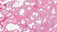
Smoking-related interstitial fibrosis (SRIF)
Smoking-related interstitial fibrosis (SRIF) is an abnormality in the lungs characterized by excessive collagen deposition within the walls of the air sacs (interstitial fibrosis). This abnormality can be seen with a microscope and diagnosed by pathologists. It is caused by cigarette smoking.[1][2]

| Smoking-related interstitial fibrosis (SRIF) | |
|---|---|
| Specialty | Pathology |
| Symptoms | Cough |
| Usual onset | Gradual |
| Causes | Smoking |
| Diagnostic method | Pathology |
| Differential diagnosis | Non-specific interstitial pneumonia (NSIP), desquamative interstitial pneumonia (DIP), respiratory bronchiolitis-interstitial lung disease (RBILD) |
| Prevention | Avoidance of cigarette smoking |
| Treatment | Smoking cessation |
| Prognosis | Stable disease over several years |
| Deaths | Not reported |
The term SRIF was coined by Dr. Anna-Luise Katzenstein (a pathologist) and colleagues in 2010 in a study of lung specimens surgically removed for lung cancer.[3] Since then, other investigators have confirmed the same abnormality in the lungs of a subset of smokers.[4][5]
Definition
The defining feature of smoking-related interstitial fibrosis is a distinctive/unique type of fibrosis characterized by "ropey" collagen bundles within the walls of the air sacs (alveoli), almost always in association with other smoking-related abnormalities such as pigmented macrophages and emphysema.[6]
Clinical symptoms
Most cases of SRIF are discovered incidentally by pathologists examining lung tissue from lungs that have been removed surgically from patients with lung cancer. These patients generally have no symptoms attributable to fibrosis, although they may have symptoms attributable to emphysema (COPD), which is a common abnormality in smokers with lung cancer. A few cases of SRIF in which patients were seen by doctors because of cough and shortness of breath have also been reported. These patients were all heavy smokers and considered to have a form of interstitial lung disease. Most were in their 40s and had abnormalities in pulmonary function tests, most commonly reduced diffusion capacity for carbon monoxide (DLCO). Their symptoms generally remained stable (did not worsen) over up to 10 years of follow-up.[7]
Role of smoking
To date, all reported patients with SRIF have been heavy current smokers or ex-smokers, lending credence to the assertion that this condition is caused by smoking.[7][3] There are no published reports of SRIF in never-smokers.
Imaging findings
Most cases of SRIF are not detectable clinically (clinically occult), and have no visible abnormalities on chest CT.[5] In clinically occult cases, a range of findings have been described on CT, including no abnormalities, low attenuation areas, clustered cysts with visible walls, and ground-glass opacities with or without reticulation.[8] In symptomatic patients, the radiologic features described on chest computed tomograms (chest CT, or CT scans of the chest) include bilateral ground-glass opacities (haziness in both lungs) and emphysema without reticulation, traction bronchiectasis or honeycombing.[7]
Treatment and Prognosis
There is very limited data on treatment and follow-up of smoking-related interstitial fibrosis. One study reported that corticosteroids were not beneficial. Smoking cessation resulted in stable/non-progressive disease after several years of follow-up. Improvements in pulmonary function tests with smoking cessation have also been documented.[7]
References
- Wick MR (September 2018). "Pathologic features of smoking-related lung diseases, with emphasis on smoking-related interstitial fibrosis and a consideration of differential diagnoses". Semin Diagn Pathol. 35 (5): 315–323. doi:10.1053/j.semdp.2018.08.002. PMID 30154023. S2CID 52111645.
- Katzenstein AL (January 2012). "Smoking-related interstitial fibrosis (SRIF), pathogenesis and treatment of usual interstitial pneumonia (UIP), and transbronchial biopsy in UIP". Mod Pathol. 25 (Suppl 1): S68–78. doi:10.1038/modpathol.2011.154. PMID 22214972.
- Katzenstein AL, Mukhopadhyay S, Zanardi C, Dexter E (March 2010). "Clinically occult interstitial fibrosis in smokers: classification and significance of a surprisingly common finding in lobectomy specimens". Hum Pathol. 41 (3): 316–25. doi:10.1016/j.humpath.2009.09.003. PMID 20004953.
- Primiani A, Dias-Santagata D, Iafrate AJ, Kradin RL (2014). "Pulmonary adenocarcinoma mutation profile in smokers with smoking-related interstitial fibrosis". International Journal of Chronic Obstructive Pulmonary Disease. 9: 525–531. doi:10.2147/COPD.S61932. PMC 4043428. PMID 24920890.
- Fabre A, Treacy A, Lavelle LP, Narski M, Faheem N, Healy D, Dodd JD, Keane MP, Egan JJ, Jebrak G, Mal H, Butler MW (December 2017). "Smoking-Related Interstitial Fibrosis: Evidence of Radiologic Regression with Advancing Age and Smoking Cessation". COPD. 14 (6): 603–609. doi:10.1080/15412555.2017.1378631. PMID 29043847. S2CID 25471885.
- Katzenstein AL (2013). "Smoking-related interstitial fibrosis (SRIF): pathologic findings and distinction from other chronic fibrosing lung diseases". Journal of Clinical Pathology. 66 (10): 882–887. doi:10.1136/jclinpath-2012-201338. PMID 23626009. S2CID 38410061.
- Vehar SJ, Yadav R, Mukhopadhyay S, Nathani A, Tolle LB (December 2022). "Smoking-Related Interstitial Fibrosis (SRIF) in Patients Presenting With Diffuse Parenchymal Lung Disease". Am J Clin Pathol. 159 (2): 146–157. doi:10.1093/ajcp/aqac144. PMC 9891418. PMID 36495281.
- Otani H, Tanaka T, Murata K, Fukuoka J, Nitta N, Nagatani Y, Sonoda A, Takahashi M (2016). "Smoking-related interstitial fibrosis combined with pulmonary emphysema: computed tomography-pathologic correlative study using lobectomy specimens". Int J Chron Obstruct Pulmon Dis. 11: 1521–32. doi:10.2147/COPD.S107938. PMC 4938241. PMID 27445472.