Solorina crocea
Solorina crocea, commonly known as the orange chocolate chip lichen, is a species of terricolous (ground-dwelling) and foliose (leafy) lichen in the family Peltigeraceae. The lichen, which was first formally described by Carl Linnaeus in 1753, has an arctic–alpine and circumpolar distribution and occurs in Asia, Europe, North America, and New Zealand. It generally grows on the bare ground in sandy soils, often in moist soil near snow patches or seepage areas. Although several forms and varieties of the lichen have been proposed in its history, these are not considered to have any independent taxonomic significance.
| Solorina crocea | |
|---|---|
.jpg.webp) | |
| In Wells Gray Provincial Park, British Columbia; scale bar is 1 cm (3⁄8 in) | |
| Scientific classification | |
| Domain: | Eukaryota |
| Kingdom: | Fungi |
| Division: | Ascomycota |
| Class: | Lecanoromycetes |
| Order: | Peltigerales |
| Family: | Peltigeraceae |
| Genus: | Solorina |
| Species: | S. crocea |
| Binomial name | |
| Solorina crocea | |
| Synonyms[1] | |
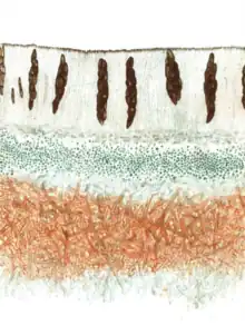
The colouration of Solorina crocea is quite distinct, making it readily identifiable: its upper thallus surface is green, while both the undersurface and its internal medulla are bright orange. The orange colour results from a pigment called solorinic acid, one of several secondary compounds that occur in the lichen. The thallus features dark brown discs, usually sunken into the surface, which are apothecia–where spores are produced. The lichen has both blue-green algae and green algae as symbiotic partners (photobionts); they are organized into separate layers in the lichen thallus.
Taxonomy
The species was one of the first lichen species formally described to science by Carl Linnaeus in his influential 1753 work Species Plantarum. He named it Lichen crocea, as it was his convention to classify all lichens in the eponymously named genus Lichen. His diagnosis made reference to the foliose thallus, the yellow underside (croceum is Latin for "saffron-coloured" or "yellow"[2]) and yellow veins on the upper side. To his understanding, the lichen occurred in Lapponia, Switzerland, and Greenland.[3] Although his 1753 work was the first official taxonomic description of the species, he had previously mentioned it in his works Flora Svecica (1745), and Flora Lapponica (1737).[4]
Later authors thought that the species was more appropriately classified in other genera, so in its taxonomic history, the taxon has been transferred to Peltigera (Georg Franz Hoffmann, 1794),[5] Peltidea (Erik Acharius, 1803),[6] Arthonia (Acharius, 1806),[7] and Parmelia (Kurt Sprengel, 1827).[8][1] The taxon is now known as the type species of Solorina,[9] a genus circumscribed by Acharius in 1808.[10]
Some forms and varieties of Solorina crocea have been described, but these do not have independent taxonomic significance, and are assessed by Index Fungorum to be synonymous with the nominate taxon. These historical subtaxa include:
- Solorina crocea f. complicata (Anzi) Bagl. & Car. (1865)
- Solorina crocea f. dubia Gyeln. (1930)[11]
- Solorina crocea f. irregularis Gyeln. (1930)[11]
- Solorina crocea var. complicata Anzi (1860)[12]
- Solorina crocea var. eutipa Gyeln. 1930[11]
Vernacular names the species has acquired in North America include the "saffron-yellow solorina",[13] and the "orange chocolate chip lichen";[14] the name refers to the large brownish fruitbodies (ascomata), which have an appearance resembling chocolate chips.[15] In the United Kingdom and Ireland, members of the genus are commonly known as "socket lichens",[16] and the species is sometimes called "mountain saffron".[17]
Description

The foliose thallus of Solorina crocea forms large rosettes comprising thick, rounded lobes;[16] it typically measures up to 6 cm (2+3⁄8 in) broad, although measurements up to 10 cm (4 in) have been recorded. It is usually tightly pressed to its growing surface.[19] The lobes comprising the thallus are 5–15 mm (3⁄16–9⁄16 in) wide with slightly upturned edges;[20] their outline varies from irregular to rounded.[14] The upper thallus surface, when moist, is olive green; in less hydrated conditions it becomes reddish brown. The lower thallus surface is bright orange with a tomentose texture and a somewhat reticulate pattern of brownish veins.[16] Lacking a cortex (an outer layer of compacted hyphae), it resembles a mat of orange-coloured, entangled threads.[18] The medulla is orange. Isidia and soredia are absent. The distinctive colouration of the upper and lower surface make this lichen readily identifiable, although in the field the colour difference may not be easy to discern if the lichen is tightly attached to its substrate.[19]
The brown to reddish-brown apothecia measure 2–10 mm (1⁄16–3⁄8 in) in diameter. They are either more or less level with the cortex or somewhat convex, rather than in concave depressions, as is the case with some other Solorina species. The photobiont partners of Solorina crocea are arranged in two distinct layers in the thallus. A more or less continuous layer with green algae lies above a patchy cyanobacterial layer.[14]
There are six to eight ascospores in each ascus of Solorina crocea. They are brown with an ellipsoid shape, and contain a single transverse septum that divides the spore into two compartments.[14] Based on a sampling of European specimens, the ascospores were measured to be an average of 11.0 μm wide (ranging from 8.4 to 21.2 μm) by 39.0 μm long (ranging from 26.6 to 51.1 μm).[21] The fine details of ascospore structure have been studied using scanning electron microscopy, revealing that there is a consistent, distinctive ornamentation in the spores of each of the most common arctic-alpine Solorina species. In S. crocea, the spores are covered with irregularly shaped papillae (small pimple-like structures), but do not form ridges or reticulations. The tips of the spores are rounded, and there is a slight constriction between the two cells.[22] Solorina octospora has similar spore size and ornamentation as S. crocea, but it does not have solorinic acid nor the subsequent orange-coloured medulla.[21]
Similar species
Solorina crocoides, described by Vilmos Kőfaragó-Gyelnik in 1930,[11] was named for its similarity with Solorina crocea. The sterile, non-fruiting version has similar chemical properties as its namesake; according to Krog and Swinscow, "The relationship between these two species needs further investigation".[23]
Some lichens collected at high elevations in Nepal are similar in overall morphology to Solorina crocea, but feature some distinguishing characteristics: they have developments of the apothecia at the lobe margins reminiscent of lichens in the genus Peltigera, there are soredia-like structures on the lobe margins that can later develop into isidia-like lobules, and they have a thick layer (100–125 μm) of cyanobacteria, while missing the green algal layer in many parts of the thallus. These lichens have not been formally described as a new species.[24]
Chemistry

Several lichen products have been found in Solorina crocea, including several anthraquinones (solorinic acid, norsolorinic acid, averantin, averantin 6-O-monomethyl ether, and 4,4'-bissolorinic acid) and two depsides (methyl gyrophorate and gyrophoric acid).[25][26] The presence of solorinic acid causes the medulla and lower thallus surface to yield a positive K spot test (K+, purple);[16] this compound may also mask the results of the KC and C reactions.[14] Solorinine is a novel amino acid that was reported from the lichen in 1994.[27] A glycoside compound extracted from S. crocea and reported in 1994 – named 1-(O-α-D-glucopyranosyl)-3S,25R-hexacosanediol – was the first of its type (a "higher alcoholic glucoside") isolated from a lichen.[28]
Three of the anthraquinones from Solorina crocea—norsolorinic acid, solorinic acid, and averantin 6-O-methyl ether—have been shown in laboratory experiments to inhibit the enzyme monoamine oxidase.[26]
Habitat and distribution
.jpg.webp)
In North America its range extends from polar regions in alpine to subalpine habitats south to California and New Mexico.[19] European countries in which the lichen has been recorded include Andorra, Austria, Bulgaria, Spain,[21] Iceland,[29] and Finland.[20] In the UK, it occurs in the Scottish Highlands, and it is represented by a single record from Ireland.[16] Its distribution in Italy is limited to the high-altitude Alps and the northern Apennines.[30] In India, it is known from the Himalayas regions (Himachal Pradesh and Sikkim).[31] Its range extends to the Eastern Himalayas, as it has been recorded from Xizang in Tibet at an altitude of 4,820 m (15,810 ft).[24] In New Zealand, it has been collected from Mount Peel at an altitude of 1,350 m (4,430 ft).[32]
Recorded habitats for the lichen include boulder fields, leached windswept ridges, and areas of solifluction.[16] In Iceland, Solorina crocea occurs in Salix herbacea snowbeds and north-facing slopes with abundant grasses and sedges.[29] Generally, the lichen occurs on the bare ground in sandy soil.[20] It grows in soils that are both acid-rich and base-rich as a result of seepage from late-lying snow patches.[13]
Species interactions
Solorina crocea is often infected by Rhagadostoma lichenicola, a lichenicolous (lichen-dwelling) fungus. Infection results in the appearance of crowded blackish ascomata on the upper surface of the host thallus and a highly branched, dark mycelium below the fruitbodies. Its presence does not seem to affect the fertility of the lichen, suggesting that the fungus requires the function and integrity of the host to remain intact. The abundance of certain strains in the associated bacterial community of the lichen has been shown to be altered by the fungal infection. Bacteria normally associated with the lichen include members of the phyla Acidobacteriota, Planctomycetota, and Proteobacteria.[15] Another lichenicolous fungus, Talpapellis solorinae, was described as a new species in 2015 from collections made in North Caucasus; it does not cause distinct damage to the infected host thallus.[33] Other fungi that have been recorded infecting Solorina crocea include Arthonia peltigerina, Cercidospora punctillata, Corticifraga peltigerae,[34] Cercidospora lichenicola, Pyrenidium actinellum,[35] Stigmidium croceae, Protothelenella croceae,[36] Thelocarpon epibolum,[37] and Pronectria robergei. Infection by Stigmidium solorinarium results in the appearance of minute perithecia, while infection by Scutula tuberculosa will result in greyish to black lecideine apothecia.[16]
Research
Researchers have cloned a gene for polyketide synthase from Solorina crocea and transferred it to the filamentous fungus Aspergillus oryzae. Lichen polyketides are of general research interest for their role in the biosynthesis of a great number of secondary metabolites.[39]
Laccases from the lichen have been purified and studied. These enzymes depolymerize and decolorize humic acids from soils, suggesting that the lichen is involved in the turnover and transformation of organic matter in the soil.[40]
A metagenome of Solorina crocea was reported in 2022. In addition to the fungal component of the lichen, the metagenome contained DNA from its green algal photobiont partner Coccomyxa solorinae, from its cyanobiont partner Nostoc, as well as from a bacterium (family Caulobacteraceae), and a Trichoderma fungus.[41]
References
- "Synonymy: Solorina crocea (L.) Ach., K. Vetensk-Acad. Nya Handl. 29: 228 (1808)". Species Fungorum. Retrieved 29 August 2022.
- Quattrocchi, Umberto (2016). CRC World Dictionary of Medicinal and Poisonous Plants: Common Names, Scientific Names, Eponyms, Synonyms, and Etymology. Boca Raton. p. 843. ISBN 978-0-429-17148-2.
{{cite book}}: CS1 maint: location missing publisher (link) - Linnaeus, Carl (1753). Species Plantarum [Species of Plants] (in Latin). Vol. 2. Stockholm: Impensis Laurentii Salvii. p. 1149.
- Jørgensen, Per M. (1994). "Linnaean lichen names and their typification". Botanical Journal of the Linnean Society. 115 (4): 261–405. doi:10.1111/j.1095-8339.1994.tb01784.x.
- Hoffmann, G.F. (1794). Descriptio et Adumbratio plantarum e classe cryptogamica Linnaei, quae Lichenes dicuntur [A description and sketch of the plants of the cryptogamic class of Linnaeus, which are called lichens] (in Latin). Vol. 2. Lepizig: Apud Siegfried Lebrecht Crusium. p. 60.
- Acharius, E. (1803). Methodus qua Omnes Detectos Lichenes: Secundum Organa Carpomorpha ad Genera, Species et Varietates Redigere atque Observationibus Illustrare Tentavit Erik Acharius (in Latin). Stockholm: Impensis F.D.D. Ulrich. p. 290.
- Acharius, Erik (1806). "Arthonia, novum genus Lichenum" [Arthonia new lichen genus]. Neues Journal für die Botanik (in Latin). 1: 1–23 [20].
- Sprengel, C. (1827). Caroli Linnaei systema vegetabilium [Charles Linnaeus' system of vegetables] (in Latin). Vol. 4. Gottingen: Sumtibus Librariae Dieterichianae. p. 280.
- "Record Details: Solorina Ach., K. Vetensk-Acad. Nya Handl. 29: 228 (1808)". Index Fungorum. Retrieved 30 September 2022.
- Acharius, Erik (1808). "Förteckning på de i Sverige våxande arter af Lafvarnas Familj" [List of the species of the Lafvarnas Family growing in Sweden]. Kongliga Vetenskaps Academiens Nya Handlingar (in Swedish). 29: 228–237.
- Gyelnik, V. (1930). "Lichenologische Mitteilungen. 20–45" [Lichenological Communications. 20–45]. Magyar Botanikai Lapok (in German). 29: 23–35.
- Anzi, Martino (1860). Catalogus lichenum: quos in provincia Sondriensi et circa Novum-Comum collegit et in ordinem systematicum digessit [A catalog of lichens: collected in the province of Sondria and around Novum-Comus and arranged in a systematic order] (in Latin). Ex officina C. Franchi. p. 24.
- Goward, Trevor; McCune, Bruce; Meidinger, Del (1994). The Lichens of British Columbia: Illustrated Keys. Part 1 — Foliose and Squamulose Species. Victoria, B.C.: Ministry of Forests Research Program. p. 125. ISBN 0-7726-2194-2. OCLC 31651418.
- Brodo, Irwin M.; Sharnoff, Sylvia Duran; Sharnoff, Stephen (2001). Lichens of North America. New Haven: Yale University Press. p. 655. ISBN 978-0-300-08249-4.
- Grube, Martin; Köberl, Martina; Lackner, Stefan; Berg, Christian; Berg, Gabriele (2012). "Host-parasite interaction and microbiome response: effects of fungal infections on the bacterial community of the Alpine lichen Solorina crocea". FEMS Microbiology Ecology. 82 (2): 472–481. doi:10.1111/j.1574-6941.2012.01425.x. PMID 22671536.
- Gilbert, O.L. (2009). "Solorina Ach. (1808)". In Smith, C.W.; Aptroot, A.; Coppins, B.J.; Fletcher, F.; Gilbert, O.L.; James, P.W.; Wolselely, P.A. (eds.). The Lichens of Great Britain and Ireland (2nd ed.). London: The Natural History Museum. pp. 844–845. ISBN 978-0-9540418-8-5.
- "Solorina crocea (L.) Ach". National Biodiversity Network Scotland. Retrieved 16 October 2022.
- Cole, Arthur C., ed. (1884). "Structure of the apothecium in Solorina". Studies in Microscopical Science. Vol. 2. London: Bailliere, Tindall and Cox. pp. 13–16.
- Geiser, Linda; McCune, Bruce (1997). Macrolichens of the Pacific Northwest (2nd ed.). Corvallis, Oregon: Oregon State University Press. p. 313. ISBN 978-0-87071-394-1.
- Stenroos, Soili; Ahti, Teuovo; Lohtander, Katileena; Myllys, Leena (2011). Suomen jäkäläopas [Finnish Lichen Guide] (in Finnish). Helsinki: Kasvimuseo, Luonnontieteellinen keskusmuseo. pp. 416–417. ISBN 978-952-10-6804-1. OCLC 767578333.
- Martínez, Isabel; Burgaz, Ana Rosa (1998). "Revision of the genus Solorina (Lichenes) in Europe based on spore size variation" (PDF). Annales Botanici Fennici. 35 (2): 137–142. JSTOR 23726541.
- Thomson, Norman F.; Thomson, John W. (1984). "Spore ornamentation in the lichen genus Solorina". The Bryologist. 87 (2): 151–153. doi:10.2307/3243122. JSTOR 3243122.
- Krog, Hildur; Swinscow, T.D.V. (1986). "Solorina sinensis and S. saccata". The Lichenologist. 18 (1): 57–62. doi:10.1017/s0024282986000075. S2CID 85934634.
- Obermayer, Walter (2004). "Additions to the lichen flora of the Tibetan region". In Döbbeler, P.; Rambold, G. (eds.). Contribution to Lichenology. Festschrift in Honour of Hannes Hertel. Bibliotheca Lichenologica. Vol. 88. Berlin/Stuttgart: J. Cramer. pp. 479–526 [508]. ISBN 978-3-443-58067-4.
- Steglich, W.; Jedtke, K.-F. (1976). "Notizen: Neue Anthrachinonfarbstoffe aus Solorina crocea" [Novel anthraquinone pigments from Solorina crocea]. Zeitschrift für Naturforschung C (in German). 31 (3–4): 197–198. doi:10.1515/znc-1976-3-420. S2CID 100627434.
- Okuyama, Emi; Hossain, Chowdhury Faiz; Yamazaki, Mikio (1991). "Monoamine oxidase inhibitors from a lichen, Solorina crocea (L.) Ach" (PDF). Shoyakugaku Zasshl. 45 (2): 159–162.
- Matsubara, Hideki; Kinoshita, Kaoru; Koyama, Kiyotaka; Takahashi, Kunio; Yoshimura, Isao; Yamamoto, Yoshikazu; Kawai, Ken-Ichi (1994). "An amino acid from Solorina crocea". Phytochemistry. 37 (4): 1209–1210. doi:10.1016/s0031-9422(00)89560-6. PMID 7765661.
- Kinoshita, Kaoru; Matsubara, Hideki; Koyama, Kiyotaka; Takahashi, Kunio; Yoshimura, Isao; Yamamoto, Yoshikazu; Miura, Yasutaka; Kinoshita, Yasuhiro; Kawai, Ken-Ichi (1994). "Topics in the chemistry of lichen compounds". Journal of the Hattori Botanical Laboratory. 76: 227–233.
- Hansen, Eric Steen (2009). "A contribution to the lichen flora of Iceland". Folia Cryptogamica Estonica. 46: 45–54.
- Nimis, Pier Luigi (2016). The Lichens of Italy. A Second Annotated Catalogue. Trieste: Edizioni Università di Trieste. p. 372. ISBN 978-88-8303-755-9.
- Awasthi, Dharani Dhar (2007). A Compendium of the Macrolichens from India, Nepal and Sri Lanka. Dehra Dun, India: Bishen Singh Mahendra Pal Singh. pp. 450–451. ISBN 978-8121106009.
- Galloway, D.J. (1976). "H. H. Allan's early collections of New Zealand lichens". New Zealand Journal of Botany. 14 (3): 225–230. doi:10.1080/0028825x.1976.10428663.
- Zhurbenko, Mikhail P.; Heuchert, Bettina; Braun, Uwe (2015). "Talpapellis solorinae sp. nov. and an updated key to the species of Talpapellis and Verrucocladosporium". Phytotaxa. 234 (2): 191–194. doi:10.11646/phytotaxa.234.2.10.
- Zhurbenko, Mikhail P. (2008). "Lichenicolous fungi from Russia, mainly from its Arctic. II" (PDF). Mycologia Balcanica. 5: 13–22.
- Zhurbenko, M.P. (2004). "Lichenicolous and some interesting lichenized fungi from the Northern Ural, Komi Republic of Russia". Herzogia. 17: 77–86.
- Diederich, Paul; Lawrey, James D.; Ertz, Damien (2018). "The 2018 classification and checklist of lichenicolous fungi, with 2000 non-lichenized, obligately lichenicolous taxa". The Bryologist. 121 (3): 340–425. doi:10.1639/0007-2745-121.3.340. S2CID 92396850.
- Spribille, Toby; Fryday, Alan M.; Pérez-Ortega, Sergio; Svensson, Måns; Tønsberg, Tor; Ekman, Stefan; Holien, Håkon; Resl, Philipp; Schneider, Kevin; Stabentheiner, Edith; Thüs, Holger; Vondrák, Jan; Sharman, Lewis (2020). "Lichens and associated fungi from Glacier Bay National Park, Alaska". The Lichenologist. 52 (2): 61–181. doi:10.1017/S0024282920000079. PMC 7398404. PMID 32788812.
- Gagunashvili, Andrey N.; Davíðsson, Snorri P.; Jónsson, Zophonías O.; Andrésson, Ólafur S. (2009). "Cloning and heterologous transcription of a polyketide synthase gene from the lichen Solorina crocea". Mycological Research. 113 (3): 354–363. doi:10.1016/j.mycres.2008.11.011. PMID 19100326.
- Lisov, Alexander; Zavarzina, Anna; Zavarzin, Alexey; Demin, Vladimir; Leontievsky, Alexey (2012). "Dimeric and monomeric laccases of soil-stabilizing lichen Solorina crocea: Purification, properties and reactions with humic acids". Soil Biology and Biochemistry. 45: 161–167. doi:10.1016/j.soilbio.2011.11.004.
- Vecherskii, Maxim V.; Khayrullin, David R.; Shadrin, Andrey M.; Lisov, Alexander V.; Zavarzina, Anna G.; Zavarzin, Alexey A.; Leontievsky, Alexey A. (2022). "Metagenomes of Lichens Solorina crocea and Peltigera canina". Microbiology Resource Announcements. 11 (1): e01000-21. doi:10.1128/mra.01000-21. PMC 8759389. PMID 34989610.
External links
 Media related to Solorina crocea at Wikimedia Commons