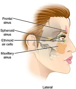
Sphenoid sinus
The sphenoid sinus is a paired paranasal sinus occurring within the body of the sphenoid bone. It represents one pair of the four paired paranasal sinuses.[1] The pair of sphenoid sinuses are separated in the middle by a septum of sphenoid sinuses. Each sphenoid sinus communicates with the nasal cavity via the opening of sphenoidal sinus.[2]: 500 The two sphenoid sinuses vary in size and shape, and are usually asymmetrical.[3]
| Sphenoid sinus | |
|---|---|
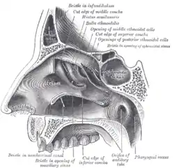 Lateral wall of nasal cavity; the three nasal conchæ have been removed. (Sphenoidal sinus visible at upper right, in dark circle.) | |
 Nose and nasal cavities. (Sphenoid sinus labeled at upper right.) | |
| Details | |
| Nerve | posterior ethmoidal nerves, and orbital branches of the pterygopalatine ganglion |
| Identifiers | |
| Latin | sinus sphenoidalis |
| MeSH | D013101 |
| TA98 | A06.1.03.003 A02.1.05.016 |
| TA2 | 600 |
| FMA | 54683 |
| Anatomical terms of bone | |
Anatomy
On average, a sphenoid sinus measures 2.2 cm vertical height, 2 cm in transverse breadth; and 2.2 cm antero-posterior depth.[3]
Each spehoid sinus is contained within the body of sphenoid bone, being situated just inferior to the sella turcica. The two sphenoid sinuses are separated medially by the septum of sphenoidal sinuses (which is usually asymmetrical).[2]: 500
An opening of sphenoidal sinus forms a passage between each sphenoidal sinus,[2]: 500 and the nasal cavity. Posteriorly, an opening of sphenoidal sinus opens into the sphenoidal sinus by an aperture high on the anterior wall the sinus; anteriorly, an opening of sphenoidal sinus opens into the roof of the nasal cavity via an aperture on the posterior wall of the sphenoethmoidal recess (occurring just superior the choana).[4]
Innervation
The mucous membrane receives sensory innervation from the posterior ethmoidal nerve (branch of the ophthalmic nerve (CN V1)), and branches of the maxillary nerve (CN V2).[5]
Postganglionic parasympathetic fibers of the facial nerve that synapsed at the pterygopalatine ganglion control mucus secretion.
Anatomical relations
Proximal structures include: the optic canal and optic nerve, internal carotid artery, cavernous sinus, trigeminal nerve, pituitary gland, and the anterior ethmoidal cells.[2]: 500
Anatomical variation
The sphenoid sinuses vary in size and shape, and, owing to the lateral displacement of the intervening septum of sphenoid sinuses, are rarely symmetrical.[3]
When exceptionally large, the sphenoid sinuses may extend into the roots of the pterygoid processes or greater wings of sphenoid bone, and may invade the basilar part of the occipital bone.[3]
The septum of the sphenoidal sinuses may be partially or completely absent. Additional incomplete septa may also be present.[2]: 500
Clinical significance
The spehnoid sinuses cannot be palpated during an extraoral examination.[1]
A potential complication of sphenoidal sinusitis is cavernous sinus thrombosis.
If a fast-growing tumor erodes the floor of the sphenoidal sinus, the vidian nerve could be in danger. If the tumor spreads laterally, the cavernous sinus and all its constituent nerves could be in danger.[6]
An endonasal surgical procedure called a sphenoidotomy may be carried out to enlarge the sphenoid sinus, usually in order to drain it.[6]
Transsphenoidal surgery
Because only thin shelves of bone separate the sphenoidal sinuses from the nasal cavities below and hypophyseal fossa above, the pituitary gland can be surgically approached through the roof of the nasal cavities by first passing through the anterioinferior aspect of the sphenoid bone and into the sinuses, followed by entry through the top of the sphenoid bone into the hypophyseal fossa.
References
![]() This article incorporates text in the public domain from page 998 of the 20th edition of Gray's Anatomy (1918)
This article incorporates text in the public domain from page 998 of the 20th edition of Gray's Anatomy (1918)
- Illustrated Anatomy of the Head and Neck, Fehrenbach and Herring, Elsevier, 2012, page 64
- Sobotta anatomy textbook. Friedrich Paulsen, Tobias M. Böckers, J. Waschke, Stephan Winkler, Katja Dalkowski, Jörg Mair. Amsterdam. 2018. ISBN 978-0-7206-7617-4. OCLC 1082911887.
{{cite book}}: CS1 maint: location missing publisher (link) CS1 maint: others (link) - Gray, Henry (1918). Gray's Anatomy (20th ed.). pp. 998–999.
- Human Anatomy, Jacob, Elsevier, 2008, page 211
- Morton, David A. (2019). The Big Picture: Gross Anatomy. K. Bo Foreman, Kurt H. Albertine (2nd ed.). New York. p. 246. ISBN 978-1-259-86264-9. OCLC 1044772257.
{{cite book}}: CS1 maint: location missing publisher (link) - Kozłowski, Z; Mazerant, M; Skóra, W; Dabrowska, K (2008). "[Sphenoidotomy--the treatment of patients with isolated sphenoid sinus diseases]". Otolaryngologia Polska = the Polish Otolaryngology. 62 (5): 582–6. doi:10.1016/S0030-6657(08)70319-6. PMID 19004262.
External links
- Anatomy photo:33:st-0712 at the SUNY Downstate Medical Center
- lesson9 at The Anatomy Lesson by Wesley Norman (Georgetown University) (latnasalwall3)