Spider toxin
Spider toxins are a family of proteins produced by spiders which function as neurotoxins. The mechanism of many spider toxins is through blockage of calcium channels.
| Spider toxin | |||||||||
|---|---|---|---|---|---|---|---|---|---|
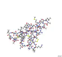
 Solution structure of omega-agatoxin-Aa4a from Agelenopsis aperta.[1] | |||||||||
| Identifiers | |||||||||
| Symbol | Toxin_9 | ||||||||
| Pfam | PF02819 | ||||||||
| Pfam clan | CL0083 | ||||||||
| InterPro | IPR004169 | ||||||||
| SCOP2 | 1oav / SCOPe / SUPFAM | ||||||||
| OPM superfamily | 112 | ||||||||
| OPM protein | 1agg | ||||||||
| |||||||||
| Delta Atracotoxin | |||||||||
|---|---|---|---|---|---|---|---|---|---|
| Identifiers | |||||||||
| Symbol | Atracotoxin | ||||||||
| Pfam | PF05353 | ||||||||
| InterPro | IPR008017 | ||||||||
| SCOP2 | 1qdp / SCOPe / SUPFAM | ||||||||
| OPM protein | 1vtx | ||||||||
| |||||||||
| Spider toxin CSTX family | |||||||||
|---|---|---|---|---|---|---|---|---|---|
| Identifiers | |||||||||
| Symbol | Toxin_35 | ||||||||
| Pfam | PF10530 | ||||||||
| InterPro | IPR011142 | ||||||||
| PROSITE | PDOC60029 | ||||||||
| |||||||||
| Spider potassium channel inhibitory toxin | |||||||||
|---|---|---|---|---|---|---|---|---|---|
| Identifiers | |||||||||
| Symbol | Toxin_12 | ||||||||
| Pfam | PF07740 | ||||||||
| Pfam clan | CL0083 | ||||||||
| InterPro | IPR011696 | ||||||||
| SCOP2 | 1d1h / SCOPe / SUPFAM | ||||||||
| OPM superfamily | 112 | ||||||||
| OPM protein | 1qk6 | ||||||||
| |||||||||
A remotely related group of atracotoxins operate by opening sodium channels. Delta atracotoxin from the venom of the Sydney funnel-web spider produces potentially fatal neurotoxic symptoms in primates by slowing the inactivation of voltage-gated sodium channels.[2] The structure of atracotoxin comprises a core beta region containing a triple-stranded a thumb-like extension protruding from the beta region and a C-terminal helix. The beta region contains a cystine knot motif, a feature seen in other neurotoxic polypeptides[2] and other spider toxins, of the CSTX family.
Spider potassium channel inhibitory toxins is another group of spider toxins. A representative of this group is hanatoxin, a 35 amino acid peptide toxin which was isolated from Chilean rose tarantula (Grammostola rosea, syn. G. spatulata) venom. It inhibits the drk1 voltage-gated potassium channel by altering the energetics of gating.[3] See also Huwentoxin-1.[4]
See also
References
- PDB: 1IVA; Reily MD, Holub KE, Gray WR, Norris TM, Adams ME (December 1994). "Structure-activity relationships for P-type calcium channel-selective omega-agatoxins". Nat. Struct. Biol. 1 (12): 853–6. doi:10.1038/nsb1294-853. PMID 7773772. S2CID 42176867.
- Mackay JP, King GF, Fletcher JI, Chapman BE, Howden ME (1997). "The structure of versutoxin (delta-atracotoxin-Hv1) provides insights into the binding of site 3 neurotoxins to the voltage-gated sodium channel". Structure. 5 (11): 1525–1535. doi:10.1016/S0969-2126(97)00301-8. PMID 9384567.
- Shimada I, Sato K, Takahashi H, Kim JI, Min HJ, Swartz KJ (2000). "Solution structure of hanatoxin1, a gating modifier of voltage-dependent K(+) channels: common surface features of gating modifier toxins". J. Mol. Biol. 297 (3): 771–780. doi:10.1006/jmbi.2000.3609. PMID 10731427.
- InterPro: IPR013140
Further reading
- Kim JI, Konishi S, Iwai H, Kohno T, Gouda H, Shimada I, Sato K, Arata Y (July 1995). "Three-dimensional solution structure of the calcium channel antagonist omega-agatoxin IVA: consensus molecular folding of calcium channel blockers". J. Mol. Biol. 250 (5): 659–71. doi:10.1006/jmbi.1995.0406. PMID 7623383.