Suillus brevipes
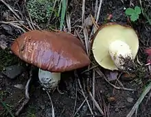
Suillus brevipes is a species of fungus in the family Suillaceae. First described by American mycologists in the late 19th century, it is commonly known as the stubby-stalk or the short-stemmed slippery Jack. The fruit bodies (mushrooms) produced by the fungus are characterized by a chocolate to reddish-brown cap covered with a sticky layer of slime, and a short whitish stipe that has neither a partial veil nor prominent, colored glandular dots. The cap can reach a diameter of about 10 cm (3+7⁄8 in), while the stipe is up to 6 cm (2+3⁄8 in) long and 2 cm (3⁄4 in) thick. Like other bolete mushrooms, S. brevipes produces spores in a vertically arranged layer of spongy tubes with openings that form a layer of small yellowish pores on the underside of the cap.
| Suillus brevipes | |
|---|---|
 | |
| Scientific classification | |
| Domain: | Eukaryota |
| Kingdom: | Fungi |
| Division: | Basidiomycota |
| Class: | Agaricomycetes |
| Order: | Boletales |
| Family: | Suillaceae |
| Genus: | Suillus |
| Species: | S. brevipes |
| Binomial name | |
| Suillus brevipes | |
| Synonyms[1] | |
| Suillus brevipes | |
|---|---|
| Pores on hymenium | |
| Cap is convex or flat | |
| Hymenium is adnate or decurrent | |
| Stipe is bare | |
| Spore print is brown | |
| Ecology is mycorrhizal | |
| Edibility is choice | |
Suillus brevipes grows in a mycorrhizal association with various species of two- and three-needled pines, especially lodgepole and ponderosa pine. The fungus is found throughout North America, and has been introduced to several other countries via transplanted pines. In the succession of mycorrhizal fungi associated with the regrowth of jack pine after clearcutting or wildfires, S. brevipes is a multi-stage fungus, found during all stages of tree development. The mushrooms are edible, and are high in the essential fatty acid linoleic acid.
Taxonomy
The species was first described scientifically as Boletus viscosus by American mycologist Charles Frost in 1874. In 1885, Charles Horton Peck, who had found specimens in pine woods of Albany County, New York, explained that the species name was a taxonomic homonym (Boletus viscosus was already in use for another species named by Ventenat in 1863[2]), and so renamed it to Boletus brevipes.[3][4] Its current name was assigned by German Otto Kuntze in 1898.[5] William Alphonso Murrill renamed it as Rostkovites brevipes in 1948;[6] the genus Rostkovites is now considered to be synonymous with Suillus.[7]
Agaricales specialist Rolf Singer included Suillus brevipes in the subsection Suillus of genus Suillus, an infrageneric (a taxonomic level below genus) grouping of species characterized by a cinnamon-brown spore print, and pores less than 1 mm wide.[8]
The specific epithet is derived from the Latin brevipes, meaning "short-footed".[9] The mushroom is commonly known as the "stubby-stalk"[10] or the "short-stemmed slippery Jack".[11]
Description
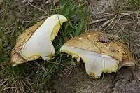
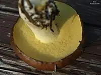
The cap is deep brown to reddish-brown, fading to tan or yellowish with age,[12] and it does not bruise with handling. The cap surface is smooth, and, depending on the moisture in the environment, may range from sticky to the touch to slimy. Depending on its maturity, the cap shape may range from spherical to broadly convex. The cap diameter measures 5–10 cm (2–3+7⁄8 in),[13] and the cap cuticle can be peeled from the surface. The tubes are yellow, becoming olive-green with age, and they have an attachment to the stipe that ranges from adnate (with most of the tube fused to the stipe) to decurrent (with the tubes broadly attached, but running somewhat down the length of the stipe). They are typically up to 1 cm (3⁄8 in) deep, and there are about 1–2 tube mouths (pores) per millimeter.[14] The pores are pale yellow, round, 1–2 mm wide, and do not change color when bruised.[15]
The stipe is white to pale yellow, dry, solid, not bruising, and pruinose (having a very fine whitish powder on the surface). A characteristic feature of many Suillus species are the glandular dots found on the stipe—clumps of hyphal cell ends through which the fungus secretes various metabolic wastes, leaving a sticky or resinous "dot". In S. brevipes, the form of the glandular dots is variable: they may be absent, slightly underdeveloped or obscurely formed with age. The stipe is usually short in comparison to the diameter of the cap, typically 2–6 cm (3⁄4–2+3⁄8 in) long and 1–2 cm (3⁄8–3⁄4 in) thick. It is either of equal width throughout, or may taper downwards; its surface bears minute puncture holes at maturity, and is it slightly fibrous at the base.[16] Collections made in New Zealand tend to have a reddish coloration at the very base of the stipe.[17] The flesh of the mushroom is initially white, but turns pale yellow in age. The odor and taste are mild. The spore print is cinnamon-brown.[18]
Microscopic characteristics
The spores are elliptical to oblong, smooth, and have dimensions of 7–10 by 3–4 µm.[15] The spore-bearing cells, the basidia, are thin-walled, club-shaped to roughly cylindrical, and measure 2–25 by 5–7 µm. They bear either two or four spores. The pleurocystidia (cystidia that are found on the face of a gill) are roughly cylindrical with rounded ends, thin-walled, and 40–55 by 5–8 µm. The cells often have brown contents, and in the presence of 2% potassium hydroxide (KOH) will appear hyaline (translucent) or vinaceous (red wine-colored); in Melzer's reagent they become pale yellow or brown. The cheilocystidia (cystidia found on the edge of a gill) are 30–60 by 7–10 µm, club-shaped to almost cylindrical, thin-walled, with brown incrusting material at the base, and arranged like a bundle of fibers. In KOH they appear hyaline, and are pale yellow in Melzer's reagent. Caulocystidia (found on the stipe) are 60–90 by 7–9 µm, mostly cylindrical with rounded ends, and arranged in bundles with brown pigment particles at the base. The caulocystidia stain vinaceous in KOH. The cuticle of the cap is made of a layer of interwoven gelatinous hyphae that are individually 2–5 µm thick; the gelatinous hyphae are responsible for the sliminess of the cuticle.[16] There are no clamp connections in the hyphae.[15]
Edibility
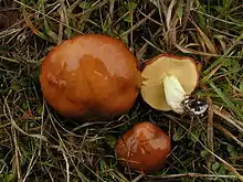
Like many species of the genus Suillus, S. brevipes is edible, and the mushroom is considered choice by some.[18][19] The odor is mild, and the taste mild or slightly acidic.[9] Field guides typically recommended to remove the slimy cap cuticle, and, in older specimens, the tube layer before consumption.[9][20] The mushrooms are common in the diet of grizzly bears in Yellowstone National Park.[21]
The fatty acid composition of S. brevipes fruit bodies has been analyzed. The cap contained a higher lipid content than the stipe—18.4% of the dry weight, compared to 12.4%. In the cap, linoleic acid made up 50.7% of the total lipids (65.7% in the stipe), oleic acid was 29.9% (12.4% in the stipe), followed by palmitic acid at 10.5% (12.6% in the stipe).[22] Linoleic acid—a member of the group of essential fatty acids called omega-6 fatty acids—is an essential dietary requirement for humans.[23]
Similar species
Several Suillus species which grow under pines could be confused with S. brevipes. S. granulatus has a longer stipe, and distinct raised granules on the stipe. S. brevipes is differentiated from S. albidipes by not having a cottony roll of velar tissue (derived from a partial veil) at the margin when young. S. pallidiceps is by distinguished its pale yellow cap color; and S. albivelatus has a veil.[15] S. pungens has a characteristic pungent odor, compared to the mild smell of S. brevipes, and like S. granulatus, has glandular dots on the stipe.[18] Boletus flaviporus is also similar.[24]
Molecular phylogenetic analyses of ribosomal DNA sequences shows that the most closely related species to S. brevipes include S. luteus, S. pseudobrevipes, and S. weaverae.[25]
Ecology
Suillus brevipes is a mycorrhizal fungus, and it develops a close symbiotic association with the roots of various tree species, especially pine. The underground mycelia form a sheath around the tree rootlets, and the fungal hyphae penetrate between the cortical cells of the root, forming ectomycorrhizae. In this way, the fungus can supply the tree with minerals, while the tree reciprocates by supplying carbohydrates created by photosynthesis. In nature, it associates with two- and three-needle pines, especially lodgepole and ponderosa pine. Under controlled laboratory conditions, the fungus has been shown to form ectomycorrhizae with ponderosa, lodgepole,[26] loblolly, eastern white,[27][28] patula,[29] pond,[30] radiata,[31] and red pines.[28] In vitro mycorrhizal associations formed with non-pine species include Pacific madrone, bearberry,[32] western larch, Sitka spruce, and coast Douglas-fir.[33] Fungal growth is inhibited by the presence of high levels of the heavy metals cadmium (350 ppm), lead (200 ppm), and nickel (20 ppm).[34]
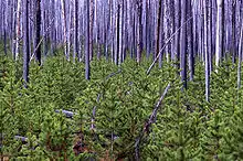
During the regrowth of pine trees after disturbance like clearcutting or wildfire, there appears an orderly sequence of mycorrhizal fungi as one species is replaced by another. A study on the ecological succession of ectomycorrhizal fungi in Canadian jack pine forests following wildfire concluded that S. brevipes is a multi-stage fungus. It appears relatively early during tree development; fruit bodies were common in 6-year-old tree stands, and the fungus colonized the highest proportion of root tips. The fungus persists throughout the life of the tree, having been found in tree stands that were 41, 65, and 122 years old. There is, however, a relative reduction in the prevalence of the fungus with increasing stand age, which may be attributed to increased competition from other fungi, and a change in habitat brought about by closure of the forest canopy.[35] Generally, S. brevipes responds favorably to silvicultural practices such as thinning and clearcutting. A 1996 study demonstrated that fruit bodies increased in abundance as the severity of disturbance increased.[36] It has been suggested that the thick-walled, wiry rhizomorphs produced by the fungus may serve as an adaptation that helps it to survive and remain viable for a period of time following disturbance.[37]
Habitat and distribution
Suillus brevipes grows singly, scattered, or in groups on the ground in late summer and autumn. A common—and sometimes abundant—mushroom, it occurs over most of North America (including Hawaii[38]),[13] south to Mexico,[39] and north to Canada.[40] This species has been found in Puerto Rico growing under planted Pinus caribaea, where it is thought to have been introduced inadvertently from North Carolina by the USDA Forest Service in 1955.[41][42] Other introductions have also occurred in exotic pine plantations in Argentina, India, New Zealand,[43][44] Japan, and Taiwan.[45]
See also
References
- "Suillus brevipes (Peck) Kuntze 1898". MycoBank. International Mycological Association. Retrieved 2015-05-22.
- "Boletus viscosus Vent. 1863". MycoBank. International Mycological Association. Retrieved 2010-09-01.
- Peck CH. (1885). "Report of the Botanist (1884)". Annual Report on the New York State Museum of Natural History. 38: 110.
- Halling RE. (1983). "Boletes described by Charles C. Frost". Mycologia. 75 (1): 70–92. doi:10.2307/3792925. JSTOR 3792925.
- Kuntze O. (1898). Revisio Generum Plantarum (in German). Vol. 3. Leipzig: A. Felix. p. 535.
- Murrill WA. (1948). "Florida boletes". Lloydia. 11: 29.
- Kirk PM, Cannon PF, Minter DW, Stalpers JA (2008). Dictionary of the Fungi (10th ed.). Wallingford, Connecticut: CAB International. p. 607. ISBN 978-0-85199-826-8.
- Singer R. (1986). The Agaricales in Modern Taxonomy (4th ed.). Koenigstein, Germany: Koeltz Scientific Books. p. 756. ISBN 978-3-87429-254-2.
- Evenson VS. (1997). Mushrooms of Colorado and the Southern Rocky Mountains. Boulder, CO: Westcliffe Publishers. p. 158. ISBN 978-1-56579-192-3.
- McKnight VB, McKnight KH (1987). A Field Guide to Mushrooms, North America. Boston, Massachusetts: Houghton Mifflin. p. plate 11. ISBN 978-0-395-91090-0.
- Arora D. (1986). Mushrooms Demystified: a Comprehensive Guide to the Fleshy Fungi. Berkeley, California: Ten Speed Press. pp. 501–502. ISBN 978-0-89815-169-5.
- Trudell, Steve; Ammirati, Joe (2009). Mushrooms of the Pacific Northwest. Timber Press Field Guides. Portland, OR: Timber Press. p. 221. ISBN 978-0-88192-935-5.
- Miller HR, Miller OK (2006). North American Mushrooms: A Field Guide to Edible and Inedible Fungi. Guilford, Connecticut: FalconGuide. p. 357. ISBN 978-0-7627-3109-1.
- Healy RA, Huffman DR, Tiffany LH, Knaphaus G (2008). Mushrooms and Other Fungi of the Midcontinental United States. Bur Oak Guide. Iowa City, Iowa: University of Iowa Press. p. 171. ISBN 978-1-58729-627-7.
- Tylukti EE. (1987). Mushrooms of Idaho and the Pacific Northwest. Vol. 2. Non-gilled Hymenomycetes. Moscow, Idaho: The University of Idaho Press. ISBN 978-0-89301-097-3.
- Grund DW, Harrison AK (1976). Nova Scotian Boletes. Lehre, Germany: J. Cramer. pp. 176–78. ISBN 978-3-7682-1062-1.
- McNabb RFR. (1968). "The Boletaceae of New Zealand". New Zealand Journal of Botany. 6 (2): 137–76 (see p. 162). doi:10.1080/0028825X.1968.10429056.

- Orr DB, Orr RT (1979). Mushrooms of Western North America. Berkeley, CA: University of California Press. pp. 88–90. ISBN 978-0-520-03656-7.
- Phillips, Roger (2010). Mushrooms and Other Fungi of North America. Buffalo, NY: Firefly Books. p. 292. ISBN 978-1-55407-651-2.
- Weber NS, Smith AH (1980). The Mushroom Hunter's Field Guide. Ann Arbor, Michigan: University of Michigan Press. p. 98. ISBN 978-0-472-85610-7.
- Mattson DJ, Podruzny SR, Haroldson MA (2002). "Consumption of fungal sporocarps by Yellowstone Grizzly Bears" (PDF). Ursus. 13: 95–103.
- Sumner JL. (1973). "The fatty acid composition of Basidiomycetes". New Zealand Journal of Botany. 11 (3): 435–42. doi:10.1080/0028825x.1973.10430293.
- Russo GL. (2009). "Dietary n-6 and n-3 polyunsaturated fatty acids: from biochemistry to clinical implications in cardiovascular prevention". Biochemical Pharmacology. 77 (6): 937–46. doi:10.1016/j.bcp.2008.10.020. PMID 19022225.
- Davis, R. Michael; Sommer, Robert; Menge, John A. (2012). Field Guide to Mushrooms of Western North America. Berkeley: University of California Press. p. 329. ISBN 978-0-520-95360-4. OCLC 797915861.
- Kretzer A, Li Y, Szaro T, Bruns TD (1996). "Internal transcribed spacer sequences from 38 recognized species of Suillus sensu lato: Phylogenetic and taxonomic implications". Mycologia. 88 (5): 776–85. doi:10.2307/3760972. JSTOR 3760972.
- Grand LF. (1968). "Conifer associated and mycorrhizal syntheses of some Pacific Northwest Suillus species". Forest Science. 14 (3): 304–12.
- Doak KB. (1934). "Fungi that produce ectotrophic mycorrhizae". Phytopathology. 24: 7.
- Palm ME, Stewart EL (1984). "In vitro synthesis of mycorrhizae between presumed specific and nonspecific Pinus + Suillus combinations". Mycologia. 76 (4): 579–600. doi:10.2307/3793215. JSTOR 3793215.
- Mohan V, Natarajan K, Ingleby K (1993). "Anatomical studies on ectomycorrhizas .2. The ectomycorrhizas produced by Amanita muscaria, Laccaria laccata and Suillus brevipes on Pinus patula". Mycorrhiza. 3 (1): 43–49. doi:10.1007/bf00213467. ISSN 0940-6360. S2CID 59942804.
- Suggs EG, Grand LF (1971). "Formation of mycorrhizae in monoxenic culture by pond pine (Pinus serotina)". Canadian Journal of Botany. 50 (5): 1003–7. doi:10.1139/b72-122.
- Morrison TM, Burr R (1973). "Physiology of the mycorrhizas of Radiata Pine". Report of Forest Research Institute for 1972, New Zealand Forest Service. 1: 19–20.
- Molina R, Trappe JM (1982). "Lack of mycorrhizal specificity by the ericaceous hosts Arbutus menzeniesii and Arctostaphylos uva-ursi". New Phytologist. 90 (3): 495–509. doi:10.1111/j.1469-8137.1982.tb04482.x.
- Molina R, Trappe JM (1982). "Patterns of ectomycorrhizal host specificity and potential among Pacific Northwest conifers and fungi". Forest Science. 28: 423–58.
- McCreight JD, Schroeder DB (1982). "Inhibition of growth of nine ectomycorrhizal fungi by cadmium, lead, and nickel in vitro". Environmental and Experimental Botany. 60 (9): 1601–5.
- Visser S. (1995). "Ectomycorrhizal fungal succession in Jack Pine stands following wildfire". New Phytologist. 129 (3): 389–401. doi:10.1111/j.1469-8137.1995.tb04309.x. JSTOR 2558393. S2CID 85061542.
- Kropp BR, Albee S (1996). "The effects of silvicultural treatments on occurrence of mycorrhizal sporocarps in a Pinus contorta forest: A preliminary study". Biological Conservation. 78 (3): 313–18. doi:10.1016/S0006-3207(96)00140-1.
- Bradbury SM, Danielson RM, Visser S (1998). "Ectomycorrhizas of regenerating stands of lodgepole pine (Pinus contorta)". Canadian Journal of Botany. 76 (2): 218–27. doi:10.1139/cjb-76-2-218.
- Hemmes DE, Desjardin D (2002). Mushrooms of Hawai'i: An Identification Guide. Berkeley, California: Ten Speed Press. p. 31. ISBN 978-1-58008-339-3.
- Cappello S, Cifuentes J (1982). "New records of the genus Suillus (Boletaceae) from Mexico". Boletin de la Sociedad Mexicana de Micologia (17): 196–206. ISSN 0085-6223.
- Bossenmaier EF. (1997). Mushrooms of the Boreal Forest. Saskatoon, Canada: University Extension Press, University of Saskatchewan. p. 60. ISBN 978-0-88880-355-9.
- Miller OK Jr, Lodge DJ, Baroni TJ (2000). "New and interesting ectomycorrhizal fungi from Puerto Rico, Mona, and Guana Islands". Mycologia. 92 (3): 558–70. doi:10.2307/3761516. JSTOR 3761516.
- Vozzo JA. (1971). "Field inoculations with mycorrhizal fungi". In Hacskaylo E (ed.). Mycorrhizae, proceedings of the first North American conference on mycorrhizae. April 1969. Forest Service Misc. Publication 1189. US Department of Agriculture. pp. 187–96.
- Read DJ. (2000). "The mycorrhizal status of Pinus". In Richardson DM (ed.). Ecology and Biogeography of Pinus. Cambridge, UK: Cambridge University Press. p. 333. ISBN 978-0-521-78910-3.
- Natarajan K, Raman N (1983). "South Indian Agaricales 20. Some mycorrhizal species". Kavaka. 11: 59–66. ISSN 0379-5179.
- Yeh K-W, Chen Z-C (1983). "Boletes of Taiwan 4" (PDF). Taiwania. 28: 122–27. Archived from the original (PDF) on 2011-07-18.
External links
 Media related to Suillus brevipes at Wikimedia Commons
 Data related to Suillus brevipes at Wikispecies