Patau syndrome
Patau syndrome is a syndrome caused by a chromosomal abnormality, in which some or all of the cells of the body contain extra genetic material from chromosome 13. The extra genetic material disrupts normal development, causing multiple and complex organ defects.
| Patau syndrome | |
|---|---|
| Other names | Trisomy 13, trisomy D, T13[1] |
 | |
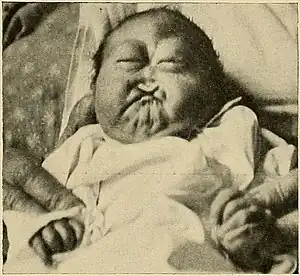
| Infant with Patau syndrome showing cleft lip, reduction of nose, and hypotelorism (c. 1912) | |
| Pronunciation |
|
| Specialty | Medical genetics |
| Usual onset | Present at birth |
| Causes | Third copy of chromosome 13 |
| Treatment | Supportive care |
| Prognosis | Poor |
| Named after | Klaus Patau |
This can occur either because each cell contains a full extra copy of chromosome 13 (a disorder known as trisomy 13 or trisomy D or T13[1]), or because each cell contains an extra partial copy of the chromosome, or because there are two different lines of cells—one healthy with the correct number of chromosomes 13 and one that contains an extra copy of the chromosome—mosaic Patau syndrome. Full trisomy 13 is caused by nondisjunction of chromosomes during meiosis (the mosaic form is caused by nondisjunction during mitosis).
Like all nondisjunction conditions (such as Down syndrome and Edwards syndrome), the risk of this syndrome in the offspring increases with maternal age at pregnancy, with about 31 years being the average.[2] Patau syndrome affects somewhere between 1 in 10,000 and 1 in 21,700 live births.[3]
Signs and symptoms


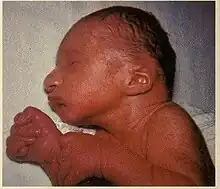
Of those fetuses that do survive to gestation and birth, common abnormalities may include:
- Nervous system
- Intellectual disability and motor disorder
- Microcephaly
- Holoprosencephaly (failure of the forebrain to divide properly) and associated facial deformities such as cyclopia
- Structural eye defects, including microphthalmia, Peters' anomaly, cataract, iris or fundus (coloboma), retinal dysplasia or retinal detachment, sensory nystagmus, cortical visual loss, and optic nerve hypoplasia
- Meningomyelocele (a spinal defect)
- Musculoskeletal and cutaneous
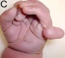
- Polydactyly (extra digits)
- Proboscis
- Congenital trigger digits
- Low-set ears[4]
- Prominent heel
- Deformed feet known as rocker-bottom feet
- Omphalocele (abdominal defect)
- Abnormal palm pattern
- Overlapping of fingers over thumb
- Cutis aplasia (missing portion of the skin/hair)
- Cleft palate
- Urogenital
- Abnormal genitalia
- Kidney defects
- Other
Causes
Patau syndrome is the result of trisomy 13, meaning each cell in the body has three copies of chromosome 13 instead of the usual two. A small percentage of cases occur when only some of the body's cells have an extra copy; such cases are called mosaic trisomy 13.
Patau syndrome can also occur when part of chromosome 13 becomes attached to another chromosome (translocated) before or at conception in a Robertsonian translocation. Affected people have two copies of chromosome 13, plus extra material from chromosome 13 attached to another chromosome. With a translocation, the person has a partial trisomy for chromosome 13 and often the physical signs of the syndrome differ from the typical Patau syndrome.
Most cases of Patau syndrome are not inherited, but occur as random events during the formation of reproductive cells (eggs and sperm). An error in cell division called non-disjunction can result in reproductive cells with an abnormal number of chromosomes. For example, an egg or sperm cell may gain an extra copy of the chromosome. If one of these atypical reproductive cells contributes to the genetic makeup of a child, the child will have an extra chromosome 13 in each of the body's cells. Mosaic Patau syndrome is also not inherited. It occurs as a random error during cell division early in fetal development.
Patau syndrome due to a translocation can be inherited. An unaffected person can carry a rearrangement of genetic material between chromosome 13 and another chromosome. This rearrangement is called a balanced translocation because there is no extra material from chromosome 13. Although they do not have signs of Patau syndrome, people who carry this type of balanced translocation are at an increased risk of having children with the condition.
Recurrence risk
Unless one of the parents is a carrier of a translocation, the chances of a couple having another trisomy 13 affected child is less than 1%, below that of Down syndrome.
Diagnosis
Diagnosis is usually based on clinical findings, although fetal chromosome testing will show trisomy 13. While many of the physical findings are similar to Edwards syndrome, there are a few unique traits, such as polydactyly. However, unlike Edwards syndrome and Down syndrome, the quad screen does not provide a reliable means of screening for this disorder. This is due to the variability of the results seen in fetuses with Patau.[6]
Treatment
Medical management of children with Trisomy 13 is planned on a case-by-case basis and depends on the individual circumstances of the patient. Treatment of Patau syndrome focuses on the particular physical problems with which each child is born. Many infants have difficulty surviving the first few days or weeks due to severe neurological problems or complex heart defects. Surgery may be necessary to repair heart defects or cleft lip and cleft palate. Physical, occupational, and speech therapy will help individuals with Patau syndrome reach their full developmental potential. Surviving children are described as happy and parents report that they enrich their lives.[7]
Prognosis
Approximately 90% of infants with Patau syndrome die within the first year of life.[8] Those children who do survive past 1 year of life are typically severely disabled with intellectual disability, seizures, and psychomotor issues. Children with the mosaic variation are usually affected to a lesser extent.[9] In a retrospective Canadian study of 174 children with trisomy 13, median survival time was 12.5 days. One and ten year survival was 19.8% and 12.9% respectively, including those who underwent aggressive surgical intervention.[10]
History
Trisomy 13 was first observed by Thomas Bartholin in 1657,[11][12] but the chromosomal nature of the disease was ascertained by Dr. Klaus Patau and Dr. Eeva Therman in 1960.[13] The disease is named in Patau's honor.
In England and Wales during 2008–09, there were 172 diagnoses of Patau syndrome (trisomy 13), with 91% of diagnoses made prenatally. There were 111 elective abortions, 14 stillbirth/miscarriage/fetal deaths, 30 outcomes unknown, and 17 live births. Approximately 4% of Patau syndrome with unknown outcomes are likely to result in a live birth, therefore the total number of live births is estimated to be 18.[14]
References
- "Trisomy 13: description in brief". GOV.UK. December 2020.
- "Prevalence and Incidence of Patau syndrome". Diseases Center-Patau Syndrome. Adviware Pty Ltd. 2008-02-04. Retrieved 2008-02-17.
mean maternal age for this abnormality is about 31 years
- About.com > Patau Syndrome (Trisomy 13) Archived 2016-03-04 at the Wayback Machine From Krissi Danielsson. Updated June 10, 2009
- H. Bruce Ostler (2004). Diseases of the eye and skin: a color atlas. Lippincott Williams & Wilkins. p. 72. ISBN 978-0-7817-4999-2. Retrieved 13 April 2010.
- "Trisomy 13: MedlinePlus Medical Encyclopedia". Retrieved 2010-04-12.
- Callahan, Tamara L., and Aaron B. Caughey. Blueprints Obstetrics & Gynecology. Baltimore, MD: Lippincott Williams & Wilkins, 2013.
- Janvier, Annie; Farlow, Wilfond (August 1, 2012). "The experience of families with children with trisomy 13 and 18 in social networks". Pediatrics. 130 (2): 293–298. doi:10.1542/peds.2012-0151. PMID 22826570. S2CID 5777839.
- Williams, G. M.; Brady, R. (2019). "Patau Syndrome". StatPearls. StatPearls. PMID 30855930.
- "Patau's Syndrome (Trisomy 13)". Archived from the original on 14 July 2014. Retrieved 3 July 2014.
- Nelson, Katherine E.; Rosella, Laura C.; Mahant, Sanjay; Guttmann, Astrid (2016). "Survival and Surgical Interventions for Children With Trisomy 13 and 18". JAMA. 316 (4): 420–8. doi:10.1001/jama.2016.9819. PMID 27458947. Retrieved 3 December 2016.
- synd/1024 at Who Named It?
- Bartholinus, Thomas (1656). Historiarum anatomicarum rariorum centuria III et IV. Ejusdem cura accessere observationes anatomicae. The Hague: Vlacq. p. 95.
- Patau K, Smith DW, Therman E, Inhorn SL, Wagner HP (1960). "Multiple congenital anomaly caused by an extra autosome". Lancet. 1 (7128): 790–3. doi:10.1016/S0140-6736(60)90676-0. PMID 14430807.
- "National Down Syndrome Cytogenetic Register Annual Reports 2008/09". Archived from the original on 2010-04-15.