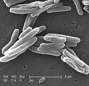
Tuberculosis verrucosa cutis
Tuberculosis verrucosa cutis is a rash of small, red papules and nodules in the skin that may appear two to four weeks after inoculation by Mycobacterium tuberculosis in a previously infected and immunocompetent individual.
| Tuberculosis verrucosa cutis | |
|---|---|
| Other names | Lupus verrucosus,[1] prosector's wart,[1] warty tuberculosis[1] anatomist's wart, verruca necrogenica |
_(14597583520).jpg.webp) | |
| Specialty | Infectious disease |
It is also known as "prosector's wart" because it was a common occupational disease of prosectors, the preparers of dissections and autopsies. Reinfection by tuberculosis via the skin, therefore, can result from accidental exposure to human tuberculous tissue in physicians, pathologists and laboratory workers; or to tissues of other infected animals, in veterinarians, butchers, etc.
TVC is one of the many forms of cutaneous tuberculosis, such as the tuberculous chancre (which results from the cutaneous inoculation in immunocompetent people without previous exposure), and the reactivation cutaneous tuberculosis (the most common form, which appears in previously infected patients). Other forms of cutaneous tuberculosis are: lupus vulgaris, scrofuloderma, lichen scrofulosorum, erythema induratum and the papulonecrotic tuberculid.
It was described by René Laennec in 1826.[2]
Signs and symptoms
Because the TVC's entry point usually is the site of a trauma, wound or puncture in the skin (during an autopsy, for example), the most frequent site for the wart are the hands. But it can occur anywhere in the skin, such as in the sole of the feet, in the anus, and, in the case of children from developing countries, in the buttocks and knees. This is because children from countries of high incidence of tuberculosis can contract the lesion after contact with tuberculous sputum, by walking barefoot, sitting or playing on the ground.
When recent, the skin lesion has the outside appearance of a wart or verruca, thus it can be confused with other kinds of warts. It evolves to an annular red-brown plaque with time, with central healing and gradual expansion in the periphery. In this phase, it can be confused with fungal infections such as blastomycosis and chromoblastomycosis.
Cause
Diagnosis
The diagnosis is confirmed by a skin biopsy and a positive culture for acid-fast bacilli. A PPD test may also result positive.
Treatment
Therapy for cutaneous tuberculosis is the same as for systemic tuberculosis, and usually consists of a 4-drug regimen, i.e., isoniazid, rifampin, pyrazinamide, and ethambutol or streptomycin.
See also
Notes
- Rapini, Ronald P.; Bolognia, Jean L.; Jorizzo, Joseph L. (2007). Dermatology: 2-Volume Set. St. Louis: Mosby. pp. Chapter 74. ISBN 978-1-4160-2999-1.
- Tigoulet F, Fournier V, Caumes E (January 2003). "[Clinical forms of the cutaneous tuberculosis]". Bull Soc Pathol Exot (in French). 96 (5): 362–7. PMID 15015840.
References
Goldman, G.; Bolognia, J.L. Pinpointing cutaneous signs of tuberculosis: is it a common wart, or tuberculosis verrucosa cutis? Archived 2004-11-16 at the Wayback Machine Journal of Critical Illness, Dec. 2002.
External links
 Media related to Tuberculosis verrucosa cutis at Wikimedia Commons
- Cutaneous tuberculosis. eMedicine.
