X-ray absorption spectroscopy
X-ray absorption spectroscopy (XAS) is a widely used technique for determining the local geometric and/or electronic structure of matter.[1] The experiment is usually performed at synchrotron radiation facilities, which provide intense and tunable X-ray beams. Samples can be in the gas phase, solutions, or solids.[2]


Background
XAS data is obtained by tuning the photon energy,[3] using a crystalline monochromator, to a range where core electrons can be excited (0.1-100 keV). The edges are, in part, named by which core electron is excited: the principal quantum numbers n = 1, 2, and 3, correspond to the K-, L-, and M-edges, respectively.[4] For instance, excitation of a 1s electron occurs at the K-edge, while excitation of a 2s or 2p electron occurs at an L-edge (Figure 1).
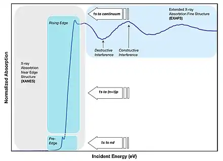
There are three main regions found on a spectrum generated by XAS data which are then thought of as separate spectroscopic techniques (Figure 2):
- The absorption threshold determined by the transition to the lowest unoccupied states:
- the states at the Fermi level in metals giving a "rising edge" with an arc tangent shape;
- the bound core excitons in insulators with a Lorentzian line-shape (they occur in a pre-edge region at energies lower than the transitions to the lowest unoccupied level);
- The X-ray absorption near-edge structure (XANES), introduced in 1980 and later in 1983 and also called NEXAFS (near-edge X-ray absorption fine structure), which are dominated by core transitions to quasi bound states (multiple scattering resonances) for photoelectrons with kinetic energy in the range from 10 to 150 eV above the chemical potential, called "shape resonances" in molecular spectra since they are due to final states of short life-time degenerate with the continuum with the Fano line-shape. In this range multi-electron excitations and many-body final states in strongly correlated systems are relevant;
- In the high kinetic energy range of the photoelectron, the scattering cross-section with neighbor atoms is weak, and the absorption spectra are dominated by EXAFS (extended X-ray absorption fine structure), where the scattering of the ejected photoelectron of neighboring atoms can be approximated by single scattering events. In 1985, it was shown that multiple scattering theory can be used to interpret both XANES and EXAFS; therefore, the experimental analysis focusing on both regions is now called XAFS.
XAS is a type of absorption spectroscopy from a core initial state with a well defined symmetry; therefore, the quantum mechanical selection rules select the symmetry of the final states in the continuum, which are usually a mixture of multiple components. The most intense features are due to electric-dipole allowed transitions (i.e. Δℓ = ± 1) to unoccupied final states. For example, the most intense features of a K-edge are due to core transitions from 1s → p-like final states, while the most intense features of the L3-edge are due to 2p → d-like final states.
XAS methodology can be broadly divided into four experimental categories that can give complementary results to each other: metal K-edge, metal L-edge, ligand K-edge, and EXAFS.
The most obvious means of mapping heterogeneous samples beyond x‐ray absorption contrast is through elemental analysis by x‐ray fluorescence, akin to EDX methods in electron microscopy.[5]
Applications
XAS is a technique used in different scientific fields including molecular and condensed matter physics,[6][7][8] materials science and engineering, chemistry, earth science, and biology. In particular, its unique sensitivity to the local structure, as compared to x-ray diffraction, have been exploited for studying:
- Amorphous solids and liquid systems
- Solid solutions
- Doping and ion implantation materials for electronics
- Local distortions of crystal lattices
- Organometallic compounds
- Metalloproteins
- Metal clusters
- Catalysis
- Vibrational dynamics[9]
- Ions in solutions
- Speciation of elements
- Liquid water and aqueous solutions
- Used to detect bone fracture
- Used to determine the concentration of any liquid in any tank
References
- "Introduction to X-Ray Absorption Fine Structure (XAFS)", X-Ray Absorption Spectroscopy for the Chemical and Materials Sciences, Chichester, UK: John Wiley & Sons, Ltd, pp. 1–8, 2017-11-24, doi:10.1002/9781118676165.ch1, ISBN 978-1-118-67616-5, retrieved 2020-09-28
- Yano J, Yachandra VK (2009-08-04). "X-ray absorption spectroscopy". Photosynthesis Research. 102 (2–3): 241–54. doi:10.1007/s11120-009-9473-8. PMC 2777224. PMID 19653117.
- Popmintchev, Dimitar; Galloway, Benjamin R.; Chen, Ming-Chang; Dollar, Franklin; Mancuso, Christopher A.; Hankla, Amelia; Miaja-Avila, Luis; O’Neil, Galen; Shaw, Justin M.; Fan, Guangyu; Ališauskas, Skirmantas (2018-03-01). "Near- and Extended-Edge X-Ray-Absorption Fine-Structure Spectroscopy Using Ultrafast Coherent High-Order Harmonic Supercontinua". Physical Review Letters. 120 (9): 093002. Bibcode:2018PhRvL.120i3002P. doi:10.1103/physrevlett.120.093002. ISSN 0031-9007. PMID 29547333.
- Kelly SD, Hesterberg D, Ravel B (2015). "Analysis of Soils and Minerals Using X-ray Absorption Spectroscopy". Methods of Soil Analysis Part 5—Mineralogical Methods. SSSA Book Series. John Wiley & Sons, Ltd. pp. 387–463. doi:10.2136/sssabookser5.5.c14. ISBN 978-0-89118-857-5. Retrieved 2020-09-24.
- Evans, John (23 November 2017). X-ray absorption spectroscopy for the chemical and materials sciences (First ed.). Hoboken, NJ. ISBN 978-1-118-67617-2. OCLC 989811256.
{{cite book}}: CS1 maint: location missing publisher (link) - Tangcharoen, T., Klysubun, W., Kongmark, C., & Pecharapa, W. (2014). Synchrotron X‐ray absorption spectroscopy and magnetic characteristics studies of metal ferrites (metal= Ni, Mn, Cu) synthesized by sol–gel auto‐combustion method. Physica Status Solidi A, 211(8), 1903-1911.https://doi.org/10.1002/pssa.201330477
- Tangcharoen, Thanit, Wantana Klysubun, and Chanapa Kongmark. "Synchrotron X-ray absorption spectroscopy and cation distribution studies of NiAl2O4, CuAl2O4, and ZnAl2O4 nanoparticles synthesized by sol-gel auto combustion method." Journal of Molecular Structure 1182 (2019): 219-229.https://doi.org/10.1016/j.molstruc.2019.01.049
- Rawat, Pankaj Singh, R. C. Srivastava, Gagan Dixit, and K. Asokan. "Structural, functional and magnetic ordering modifications in graphene oxide and graphite by 100 MeV gold ion irradiation." Vacuum 182 (2020): 109700.https://doi.org/10.1016/j.vacuum.2020.109700
- Ljungberg, Mathias (December 2017). "Vibrational effects in x-ray absorption and resonant inelastic x-ray scattering using a semiclassical scheme". Physical Review B. 96 (21): 214302. arXiv:1709.06786. Bibcode:2017PhRvB..96u4302L. doi:10.1103/PhysRevB.96.214302. S2CID 119210376. Retrieved 21 April 2023.
External links
- Zhang M (15 August 2020). "XANES - Theory". LibreTexts Project.
- Newville M (25 July 2008). "Fundamentals of XAFS" (PDF). Chicago, IL: University of Chicago.