Color constancy
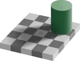
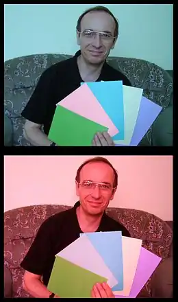
Color constancy is an example of subjective constancy and a feature of the human color perception system which ensures that the perceived color of objects remains relatively constant under varying illumination conditions. A green apple for instance looks green to us at midday, when the main illumination is white sunlight, and also at sunset, when the main illumination is red. This helps us identify objects.


Color vision
Color vision is how we perceive the objective color, which people, animals and machines are able to distinguish objects based on the different wavelengths of light reflected, transmitted, or emitted by the object. In humans, light is detected by the eye using two types of photoreceptors, cones and rods, which send signals to the visual cortex, which in turn processes those colors into a subjective perception. Color constancy is a process that allows the brain to recognize a familiar object as being a consistent color regardless of the amount or wavelengths of light reflecting from it at a given moment.[1][2]
Object illuminance
The phenomenon of color constancy occurs when the source of illumination is not directly known.[3] It is for this reason that color constancy takes a greater effect on days with sun and clear sky as opposed to days that are overcast.[3] Even when the sun is visible, color constancy may affect color perception. This is due to an ignorance of all possible sources of illumination. Although an object may reflect multiple sources of light into the eye, color constancy causes objective identities to remain constant.[4]
D. H. Foster (2011) states, "in the natural environment, the source itself may not be well defined in that the illumination at a particular point in a scene is usually a complex mixture of direct and indirect [light] distributed over a range of incident angles, in turn modified by local occlusion and mutual reflection, all of which may vary with time and position."[3] The wide spectrum of possible illuminances in the natural environment and the limited ability of the human eye to perceive color means that color constancy plays a functional role in daily perception. Color constancy allows for humans to interact with the world in a consistent or veridical manner[5] and it allows for one to more effectively make judgements on the time of day.[4][6]
Physiological basis
The physiological basis for color constancy is thought to involve specialized neurons in the primary visual cortex that compute local ratios of cone activity, which is the same calculation that Land's retinex algorithm uses to achieve color constancy. These specialized cells are called double-opponent cells because they compute both color opponency and spatial opponency. Double-opponent cells were first described by Nigel Daw in the goldfish retina.[7][8] There was considerable debate about the existence of these cells in the primate visual system; their existence was eventually proven using reverse-correlation receptive field mapping and special stimuli that selectively activate single cone classes at a time, so-called "cone-isolating" stimuli.[9][10] Human brain imaging evidence strongly suggests that a critical cortical locus for generating color constancy is located in cortical area V4,[11] damage in which leads to the syndrome of cerebral achromatopsia.
Color constancy works only if the incident illumination contains a range of wavelengths. The different cone cells of the eye register different but overlapping ranges of wavelengths of the light reflected by every object in the scene. From this information, the visual system attempts to determine the approximate composition of the illuminating light. This illumination is then discounted[12] in order to obtain the object's "true color" or reflectance: the wavelengths of light the object reflects. This reflectance then largely determines the perceived color.
Neural mechanism
There are two possible mechanisms for color constancy. The first mechanism is unconscious inference.[13] The second view holds this phenomenon to be caused by sensory adaptation.[14][15] Research suggests color constancy to be related changes in retinal cells as well as cortical areas related to vision.[16][17][18] This phenomenon is most likely attributed to changes in various levels of the visual system.[3]
Cone adaptation
Cones, specialized cells within the retina, will adjust relative to light levels within the local environment.[18] This occurs at the level of individual neurons.[19] However, this adaptation is incomplete.[3] Chromatic adaptation is also regulated by processes within the brain. Research in monkeys suggest that changes in chromatic sensitivity is correlated to activity in parvocellular lateral geniculate neurons.[20][21] Color constancy may be both attributed to localized changes in individual retinal cells or to higher level neural processes within the brain.[19]
Metamerism
Metamerism, the perceiving of colors within two separate scenes, can help to inform research regarding color constancy.[22][23] Research suggests that when competing chromatic stimuli are presented, spatial comparisons must be completed early in the visual system. For example, when subjects are presented stimuli in a dichoptic fashion, an array of colors and a void color, such as grey, and are told to focus on a specific color of the array, the void color appears different than when perceived in a binocular fashion.[24] This means that color judgements, as they relate to spatial comparisons, must be completed at or prior to the V1 monocular neurons.[24][25][26] If spatial comparisons occur later in the visual system such as in cortical area V4, the brain would be able to perceive both the color and void color as though they were seen in a binocular fashion.
Retinex theory
The "Land effect" is the capacity to see full color (if muted) images solely by looking at a photo with red and gray wavelengths. The effect was discovered by Edwin H. Land, who was attempting to reconstruct James Clerk Maxwell's early experiments in full-colored images. Land realized that, even when there were no green or blue wavelengths present in an image, the visual system would still perceive them as green or blue by discounting the red illumination. Land described this effect in a 1959 article in Scientific American.[27] In 1977, Land wrote another Scientific American article that formulated his "retinex theory" to explain the Land effect. The word "retinex" is a blend of "retina" and "cortex", suggesting that both the eye and the brain are involved in the processing. Land, with John McCann, also developed a computer program designed to imitate the retinex processes taking place in human physiology.[28]
The effect can be experimentally demonstrated as follows. A display called a "Mondrian" (after Piet Mondrian whose paintings are similar) consisting of numerous colored patches is shown to a person. The display is illuminated by three white lights, one projected through a red filter, one projected through a green filter, and one projected through a blue filter. The person is asked to adjust the intensity of the lights so that a particular patch in the display appears white. The experimenter then measures the intensities of red, green, and blue light reflected from this white-appearing patch. Then the experimenter asks the person to identify the color of a neighboring patch, which, for example, appears green. Then the experimenter adjusts the lights so that the intensities of red, blue, and green light reflected from the green patch are the same as were originally measured from the white patch. The person shows color constancy in that the green patch continues to appear green, the white patch continues to appear white, and all the remaining patches continue to have their original colors.
Color constancy is a desirable feature of computer vision, and many algorithms have been developed for this purpose. These include several retinex algorithms.[29][30][31][32] These algorithms receive as input the red/green/blue values of each pixel of the image and attempt to estimate the reflectances of each point. One such algorithm operates as follows: the maximal red value rmax of all pixels is determined, and also the maximal green value gmax and the maximal blue value bmax. Assuming that the scene contains objects which reflect all red light, and (other) objects which reflect all green light and still others which reflect all blue light, one can then deduce that the illuminating light source is described by (rmax, gmax, bmax). For each pixel with values (r, g, b) its reflectance is estimated as (r/rmax, g/gmax, b/bmax). The original retinex algorithm proposed by Land and McCann uses a localized version of this principle.[33][34]
Although retinex models are still widely used in computer vision, actual human color perception has been shown to be more complex.[35]
See also
- Chromatic adaptation
- Memory color effect
- Shadow and highlight enhancement
- Trichromacy
- Tetrachromacy
- Theory of Colours[36]
References
- Krantz, John (2009). Experiencing Sensation and Perception (PDF). Pearson Education, Limited. pp. 9.9–9.10. ISBN 978-0-13-097793-9. Archived from the original (PDF) on November 17, 2017. Retrieved January 23, 2012.
- "Wendy Carlos ColorVision1".
- Foster, David H. (2011). "Color Constancy". Vision Research. 51 (7): 674–700. doi:10.1016/j.visres.2010.09.006. PMID 20849875. S2CID 1399339.
- Jameson, D.; Hurvich, L. M. (1989). "Essay concerning color constancy". Annual Review of Psychology. 40: 1–22. doi:10.1146/annurev.psych.40.1.1. PMID 2648972.
- Zeki, S. (1993). A vision of the brain. Oxford: Blackwell Science Ltd.
- Reeves, A (1992). "Areas of ignorance and confusion in color science". Behavioral and Brain Sciences. 15: 49–50. doi:10.1017/s0140525x00067510.
- Daw, Nigel W. (November 17, 1967). "Goldfish Retina: Organization for Simultaneous Colour Contrast". Science. 158 (3803): 942–4. Bibcode:1967Sci...158..942D. doi:10.1126/science.158.3803.942. PMID 6054169. S2CID 1108881.
- Bevil R. Conway (2002). Neural Mechanisms of Color Vision: Double-Opponent Cells in the Visual Cortex. Springer. ISBN 978-1-4020-7092-1.
- Conway, BR; Livingstone, MS (2006). "Spatial and Temporal Properties of Cone Signals in Alert Macaque Primary Visual Cortex (V1)". Journal of Neuroscience. 26 (42): 10826–46. doi:10.1523/jneurosci.2091-06.2006. PMC 2963176. PMID 17050721. [cover illustration].
- Conway, BR (2001). "Spatial structure of cone inputs to color cells in alert macaque primary visual cortex (V-1)". Journal of Neuroscience. 21 (8): 2768–2783. doi:10.1523/JNEUROSCI.21-08-02768.2001. PMC 6762533. PMID 11306629. [cover illustration].
- Bartels, A.; Zeki, S. (2000). "The architecture of the colour centre in the human visual brain: new results and a review *". European Journal of Neuroscience. 12 (1): 172–193. doi:10.1046/j.1460-9568.2000.00905.x. ISSN 1460-9568. PMID 10651872. S2CID 6787155.
- "Discounting the illuminant" is a term introduced by Helmholtz: McCann, John J. (March 2005). "Do humans discount the illuminant?". In Bernice E. Rogowitz; Thrasyvoulos N. Pappas; Scott J. Daly (eds.). Proceedings of SPIE. Human Vision and Electronic Imaging X. Vol. 5666. pp. 9–16. doi:10.1117/12.594383.
- Judd, D. B. (1940). "Hue saturation and lightness of surface colors with chromatic illumination". Journal of the Optical Society of America. 30: 2–32. doi:10.1364/JOSA.30.000002.
- Helson, H (1943). "Some factors and implications of color constancy". Journal of the Optical Society of America. 33 (10): 555–567. Bibcode:1943JOSA...33..555H. doi:10.1364/josa.33.000555.
- Hering, E. (1920). Grundzüge der Lehre vom Lichtsinn. Berlin: Springer (Trans. Hurvich, L. M. & Jameson, D., 1964, Outlines of a theory of the light sense, Cambridge MA: Harvard University Press).
- Zeki, S (1980). "The representation of colours in the cerebral cortex". Nature. 284 (5755): 412–418. Bibcode:1980Natur.284..412Z. doi:10.1038/284412a0. PMID 6767195. S2CID 4310049.
- Zeki, S (1983). "Colour coding in the cerebral cortex: The reaction of cells in monkey visual cortex to wavelengths and colours". Neuroscience. 9 (4): 741–765. doi:10.1016/0306-4522(83)90265-8. PMID 6621877. S2CID 21352625.
- Hood, D.C. (1998). "Lower-Level Visual Processing and Models of Light Adaptation". Annual Review of Psychology. 49: 503–535. doi:10.1146/annurev.psych.49.1.503. PMID 9496631. S2CID 12490019.
- Lee, B. B.; Dacey, D. M.; Smith, V. C.; Pokorny, J. (1999). "Horizontal cells reveal cone type-specific adaptation in primate retina". Proceedings of the National Academy of Sciences of the United States of America. 96 (25): 14611–14616. Bibcode:1999PNAS...9614611L. doi:10.1073/pnas.96.25.14611. PMC 24484. PMID 10588753.
- Creutzfeldt, O. D.; Crook, J. M.; Kastner, S.; Li, C.-Y.; Pei, X. (1991). "The neurophysiological correlates of colour and brightness contrast in lateral geniculate neurons: 1. Population analysis". Experimental Brain Research. 87 (1): 3–21. doi:10.1007/bf00228503. PMID 1756832. S2CID 1363735.
- Creutzfeldt, O. D.; Kastner, S.; Pei, X.; Valberg, A. (1991). "The neurophysiological correlates of colour and brightness contrast in lateral geniculate neurons: II. Adaptation and surround effects". Experimental Brain Research. 87 (1): 22–45. doi:10.1007/bf00228504. PMID 1756829. S2CID 75794.
- Kalderon, Mark Eli (2008). "Metamerism, Constancy, and Knowing Which" (PDF). Mind. 117 (468): 935–971. doi:10.1093/mind/fzn043. JSTOR 20532701.
- Gupte, Vilas (December 1, 2009). "Color Constancy, by Marc Ebner (Wiley; 2007) pp 394 ISBN 978-0-470-05829-9 (HB)". Coloration Technology. 125 (6): 366–367. doi:10.1111/j.1478-4408.2009.00219.x. ISSN 1478-4408.
- Moutoussis, K.; Zeki, S. (2000). "A psychophysical dissection of the brain sites involved in color-generating comparisons". Proceedings of the National Academy of Sciences of the United States of America. 97 (14): 8069–8074. Bibcode:2000PNAS...97.8069M. doi:10.1073/pnas.110570897. PMC 16671. PMID 10859348.
- Hurlbert, A. C.; Bramwell, D. I.; Heywood, C.; Cowey, A. (1998). "Discrimination of cone contrast changes as evidence for colour constancy in cerebral achromatopsia". Experimental Brain Research. 123 (1–2): 136–144. doi:10.1007/s002210050554. PMID 9835402. S2CID 1645601.
- Kentridge, R. W.; Heywood, C. A.; Cowey, A. (2004). "Chromatic edges, surfaces and constancies in cerebral achromatopsia". Neuropsychologia. 42 (6): 821–830. doi:10.1016/j.neuropsychologia.2003.11.002. PMID 15037060. S2CID 16183218.
- Land, Edwin (May 1959). "Experiments in Color Vision" (PDF). Scientific American. 200 (5): 84-94 passim. Bibcode:1959SciAm.200e..84L. doi:10.1038/scientificamerican0559-84. PMID 13646648.
- Land, Edwin (December 1977). "The Retinex Theory of Color Vision" (PDF). Scientific American. 237 (6): 108–28. Bibcode:1977SciAm.237f.108L. doi:10.1038/scientificamerican1277-108. PMID 929159.
- Morel, Jean-Michel; Petro, Ana B.; Sbert, Catalina (2009). Eschbach, Reiner; Marcu, Gabriel G; Tominaga, Shoji; Rizzi, Alessandro (eds.). "Fast implementation of color constancy algorithms". Color Imaging XIV: Displaying, Processing, Hardcopy, and Applications. 7241: 724106. Bibcode:2009SPIE.7241E..06M. CiteSeerX 10.1.1.550.4746. doi:10.1117/12.805474. S2CID 19950750.
- Kimmel, R.; Elad, M.; Shaked, D.; Keshet, R.; Sobel, I. (2003). "A Variational Framework for Retinex" (PDF). International Journal of Computer Vision. 52 (1): 7–23. doi:10.1023/A:1022314423998. S2CID 14479403.
- Barghout, Lauren, and Lawrence Lee. Perceptual information processing system. U.S. Patent Application 10/618,543. http://www.google.com/patents/US20040059754
- Barghout, Lauren. "Visual Taxometric Approach to Image Segmentation Using Fuzzy-Spatial Taxon Cut Yields Contextually Relevant Regions." Information Processing and Management of Uncertainty in Knowledge-Based Systems. Springer International Publishing, 2014.
- Provenzi, Edoardo; De Carli, Luca; Rizzi, Alessandro; Marini, Daniele (2005). "Mathematical definition and analysis of the Retinex algorithm". JOSA A. 22 (12): 2613–2621. Bibcode:2005JOSAA..22.2613P. doi:10.1364/josaa.22.002613. PMID 16396021.
- Bertalmío, Marcelo; Caselles, Vicent; Provenzi, Edoardo (2009). "Issues About Retinex Theory and Contrast Enhancement". International Journal of Computer Vision. 83: 101–119. doi:10.1007/s11263-009-0221-5. S2CID 4613179.
- Hurlbert, A.C.; Wolf, K. The contribution of local and global cone-contrasts to colour appearance: a Retinex-like model. In: Proceedings of the SPIE 2002, San Jose, CA
- Ribe, N.; Steinle, F. (2002). "Exploratory Experimentation: Goethe, Land, and Color Theory". Physics Today. 55 (7): 43. Bibcode:2002PhT....55g..43R. doi:10.1063/1.1506750.
Retinex
Here "Reprinted in McCann" refers to McCann, M., ed. 1993. Edwin H. Land's Essays. Springfield, Va.: Society for Imaging Science and Technology.
- (1964) "The retinex" Am. Sci. 52(2): 247–64. Reprinted in McCann, vol. III, pp. 53–60. Based on acceptance address for William Procter Prize for Scientific Achievement, Cleveland, Ohio, December 30, 1963.
- with L.C. Farney and M.M. Morse. (1971) "Solubilization by incipient development" Photogr. Sci. Eng. 15(1):4–20. Reprinted in McCann, vol. I, pp. 157–73. Based on lecture in Boston, June 13, 1968.
- with J.J. McCann. (1971) "Lightness and retinex theory" J. Opt. Soc. Am. 61(1):1–11. Reprinted in McCann, vol. III, pp. 73–84. Based on the Ives Medal lecture, October 13, 1967.
- (1974) "The retinex theory of colour vision" Proc. R. Inst. Gt. Brit. 47:23–58. Reprinted in McCann, vol. III, pp. 95–112. Based on Friday evening discourse, November 2, 1973.
- (1977) "The retinex theory of color vision" Sci. Am. 237:108-28. Reprinted in McCann, vol. III, pp. 125–42.
- with H.G. Rogers and V.K. Walworth. (1977) "One-step photography" In Neblette's Handbook of Photography and Reprography, Materials, Processes and Systems, 7th ed., J. M. Sturge, ed., pp. 259–330. New York: Reinhold. Reprinted in McCann, vol. I, pp. 205–63.
- (1978) "Our 'polar partnership' with the world around us: Discoveries about our mechanisms of perception are dissolving the imagined partition between mind and matter" Harv. Mag. 80:23–25. Reprinted in McCann, vol. III, pp. 151–54.
- with D.H. Hubel, M.S. Livingstone, S.H. Perry, and M.M. Burns. (1983) "Colour-generating interactions across the corpus callosum" Nature 303(5918):616-18. Reprinted in McCann, vol. III, pp. 155–58.
- (1983) "Recent advances in retinex theory and some implications for cortical computations: Color vision and the natural images" Proc. Natl. Acad. Sci. U. S. A. 80:5136–69. Reprinted in McCann, vol. III, pp. 159–66.
- (1986) "An alternative technique for the computation of the designator in the retinex theory of color vision" Proc. Natl. Acad. Sci. U. S. A. 83:3078–80.