Dorsal interossei of the foot
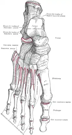
In human anatomy, the dorsal interossei of the foot are four muscles situated between the metatarsal bones.
| Dorsal interossei muscles | |
|---|---|
 The Interossei dorsales. Left foot. | |
| Details | |
| Origin | metatarsals |
| Insertion | proximal phalanges |
| Nerve | lateral plantar nerve |
| Actions | abduct toes |
| Antagonist | Plantar interossei muscles |
| Identifiers | |
| Latin | Musculi interossei dorsales pedis |
| TA98 | A04.7.02.070 |
| TA2 | 2686 |
| FMA | 37457 |
| Anatomical terms of muscle | |
Origin
The four interossei muscles are bipenniform muscles each originating by two heads from the proximal half of the sides of adjacent metatarsal bones. [1]
Insertion
The two heads of each muscle form a central tendon which passes forwards deep to the deep transverse metatarsal ligament.[1] The tendons are inserted on the bases of the second, third, and fourth proximal phalanges[2] and into the aponeurosis of the tendons of the extensor digitorum longus[3] without attaching to the extensor hoods of the toes.[1]
Thus, the first is inserted into the medial side of the second toe; the other three are inserted into the lateral sides of the second, third, and fourth toes.[3]
Action
The dorsal interossei abduct at the metatarsophalangeal joints of the third and fourth toes. Because there is a pair of dorsal interossei muscles attached on both sides of the second toe, simultaneous contraction of these muscles results in no movement. This arrangement of dorsal interossei makes the second toe the midline of the foot, whereas the midline of the hand (marked by dorsal interossei of hand) is in the third finger. [2]
Abduction is of little importance in the foot, but, together with the plantar interossei, the dorsal interossei also produce flexion at the metatarsophalangeal joints. Although small, the dorsal interossei are powerful muscles that, together with their plantar counterparts, control the direction of the toes during violent activity, thus allowing the long and short flexors to perform their actions. [1]
Because of the relationship to the metatarsophalangeal joints, the interossei muscles also contribute to maintaining the anterior metatarsal arch of the foot and also, to a limited extent, the medial and lateral longitudinal arches of the foot. [1]
Innervation
All dorsal interossei are innervated by the lateral plantar nerve (S2–3). Those in the fourth interosseous space are innervated by the superficial branch and the other by the deep branch. [1] The first and second dorsal interossei muscles additionally receive innervation from the lateral branch of the deep fibular nerve.[4]
Relations
In the angular interval left between the heads of each of the three lateral muscles, one of the perforating arteries passes to the dorsum of the foot; through the space between the heads of the first muscle the deep plantar branch of the dorsalis pedis artery enters the sole of the foot. [3]
See also
- Interosseous muscles of the hand
- Interosseous muscles of the foot
Additional images
 Bones of the right foot. Dorsal surface.
Notes
![]() This article incorporates text in the public domain from page 495 of the 20th edition of Gray's Anatomy (1918)
This article incorporates text in the public domain from page 495 of the 20th edition of Gray's Anatomy (1918)
- Palastanga, Field & Soames 2006, p. 311
- Behnke 2006, p. 227
- Gray's Anatomy, 1918 (see infobox)
- Drake, Richard (2015). Gray's Anatomy for Students. Elsevier. ISBN 978-0702051333.
References
- Behnke, Robert S. (2006). Kinetic anatomy (2nd ed.). Human Kinetics. p. 227. ISBN 0-7360-5909-1.
- Palastanga, Nigel; Field, Derek; Soames, Roger (2006). Anatomy and human movement: structure and function (5th ed.). Elsevier Health Sciences. ISBN 978-0-7506-8814-7.