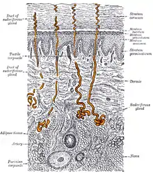
Eccrine sweat gland
Eccrine sweat glands (/ˈɛkrən, -ˌkraɪn, -ˌkriːn/; from Greek ekkrinein 'secrete';[3] sometimes called merocrine glands) are the major sweat glands of the human body,[4] found in virtually all skin, with the highest density in palm and soles, then on the head, but much less on the torso and the extremities. In other mammals, they are relatively sparse, being found mainly on hairless areas such as foot pads. They reach their peak of development in humans, where they may number 200–400/cm2 of skin surface.[5][6] They produce a clear, odorless substance, sweat, consisting primarily of water. These are present from birth. Their secretory part is present deep inside the dermis.
| Eccrine sweat gland | |
|---|---|
 A sectional view of the skin (magnified), with eccrine glands highlighted. | |
| Details | |
| Precursor | Ectoderm[1] |
| System | Integumentary[1] |
| Nerve | Cholinergic sympathetic nerves[2] |
| Identifiers | |
| Latin | Glandula sudorifera merocrina; Glandula sudorifera eccrina |
| MeSH | D004439 |
| TH | H3.12.00.3.03009 |
| FMA | 59154 |
| Anatomical terminology | |
Eccrine glands are composed of an intraepidermal spiral duct, the "acrosyringium"; a dermal duct, consisting of a straight and coiled portion; and a secretory tubule, coiled deep in the dermis or hypodermis.[7] The eccrine gland opens out through the sweat pore. The coiled portion is formed by two concentric layer of columnar or cuboidal epithelial cells.[8] The epithelial cells are interposed by the myoepithelial cells. Myoepithelial cells support the secretory epithelial cells. The duct of eccrine gland is formed by two layers of cuboidal epithelial cells.[9]
Eccrine glands are active in thermoregulation by providing cooling from water evaporation of sweat secreted by the glands on the body surface and emotional induced sweating (anxiety, fear, stress, and pain).[6][7] The white sediment in otherwise colorless eccrine secretions is caused by evaporation that increases the concentration of salts.
The odour from sweat is due to bacterial activity on the secretions of the apocrine sweat glands, a distinctly different type of sweat gland found in human skin.
Eccrine glands are the only sweat glands innervated by the parasympathetic nervous system, primarily by cholinergic fibers whose discharge is altered primarily by changes in deep body temperature (core temperature), but by adrenergic fibers of the sympathetic nervous system, as well.[10] The glands on palms and soles do not respond to temperature but secrete at times of emotional stress.
Secretion
The secretion of eccrine glands is a sterile, dilute electrolyte solution with primary components of bicarbonate, potassium, and sodium chloride (NaCl),[6] and other minor components such as glucose, pyruvate, lactate, cytokines, immunoglobulins, antimicrobial peptides (e.g., dermcidin), and many others.[6]
Relative to the plasma and extracellular fluid, the concentration of Na+ ions is much lower in sweat (~40 mM in sweat versus ~150 mM in plasma and extracellular fluid). Initially, within the eccrine glands, sweat has a high concentration of Na+ ions. The Na+ ions are re-absorbed into the tissue via the epithelial sodium channels (ENaC) that are located on the apical membrane of the cells that form the eccrine gland ducts (see Fig. 9 and Fig. 10 of the reference).[9] This re-uptake of Na+ ions reduces the loss of Na+ during the process of perspiration. Patients with the systemic pseudohypoaldosteronism syndrome who carry mutations in the ENaC subunit genes have salty sweat as they cannot reabsorb the salt in sweat.[11][12] In these patients, Na+ ion concentrations can greatly increase (up to 180 mmol/L).[11] [13]
In people who have hyperhidrosis, the sweat glands (eccrine glands in particular) overreact to stimuli and are just generally overactive, producing more sweat than normal. Similarly, cystic fibrosis patients also produce salty sweat. But in these cases, the problem is in the CFTR chloride transporter that is also located on the apical membrane of eccrine gland ducts.[9]
Dermicidin is a newly isolated antimicrobial peptide produced by the eccrine sweat glands.[14]
References
- Neas, John F. "Development of the Integumentary System". In Martini, Frederic H.; Timmons, Michael J.; Tallitsch, Bob (eds.). Embryology Atlas (4th ed.). Benjamin Cumings.
- Krstic, Radivoj V. (18 March 2004). Human Microscopic Anatomy: An Atlas for Students of Medicine and Biology. Springer. p. 464. ISBN 9783540536666.
- McKean, Erin (2005). "eccrine". The New Oxford American Dictionary (2 ed.). ISBN 9780195170771.
- "our weird lack of hair may be the key to our success".
- James, William; Berger, Timothy; Elston, Dirk (2005). Andrews' Diseases of the Skin: Clinical Dermatology (10th ed.). Saunders. pp. 6–7. ISBN 978-0-7216-2921-6.
- Bolognia, J., Jorizzo, J., & Schaffer, J. (2012). Dermatology (3rd ed., pp. 539-544). [Philadelphia]: Elsevier Saunders.
- Wilke, K.; Martin, A.; Terstegen, L.; Biel, S. S. (June 2007). "A short history of sweat gland biology". International Journal of Cosmetic Science. 29 (3): 169–179. doi:10.1111/j.1467-2494.2007.00387.x. ISSN 1468-2494. PMID 18489347. S2CID 205556581.
- Cui, Chang-Yi; Schlessinger, David (2015). "Eccrine sweat gland development and sweat secretion". Experimental Dermatology. 24 (9): 644–650. doi:10.1111/exd.12773. ISSN 0906-6705. PMC 5508982. PMID 26014472.
- Hanukoglu I, Boggula VR, Vaknine H, Sharma S, Kleyman T, Hanukoglu A (January 2017). "Expression of epithelial sodium channel (ENaC) and CFTR in the human epidermis and epidermal appendages". Histochemistry and Cell Biology. 147 (6): 733–748. doi:10.1007/s00418-016-1535-3. PMID 28130590. S2CID 8504408.
- Sokolov, VE; Shabadash, SA; Zelikina, TI (1980). "Innervation of eccrine sweat glands". Biology Bulletin of the Academy of Sciences of the USSR. 7 (5): 331–46. PMID 7317512.
- Hanukoglu A (Nov 1991). "Type I pseudohypoaldosteronism includes two clinically and genetically distinct entities with either renal or multiple target organ defects". The Journal of Clinical Endocrinology and Metabolism. 73 (5): 936–44. doi:10.1210/jcem-73-5-936. PMID 1939532.
- Hanukoglu I, Hanukoglu A (Jan 2016). "Epithelial sodium channel (ENaC) family: Phylogeny, structure-function, tissue distribution, and associated inherited diseases". Gene. 579 (2): 95–132. doi:10.1016/j.gene.2015.12.061. PMC 4756657. PMID 26772908.
- Edelheit, Oded; Hanukoglu, Israel; Shriki, Yafit; Tfilin, Matanel; Dascal, Nathan; Gillis, David; Hanukoglu, Aaron (2010). "Truncated beta epithelial sodium channel (ENaC) subunits responsible for multi-system pseudohypoaldosteronism (PHA) support partial activity of ENaC". The Journal of Steroid Biochemistry and Molecular Biology. 119 (1–2): 84–88. doi:10.1016/j.jsbmb.2010.01.002. PMID 20064610. S2CID 9564777.
- Niyonsaba, F; Suzuki, A; Ushio, H; Nagaoka, I; Ogawa, H; Okumura, K (2009). "The human antimicrobial peptide dermcidin activates normal human keratinocytes". The British Journal of Dermatology. 160 (2): 243–9. doi:10.1111/j.1365-2133.2008.08925.x. PMID 19014393. S2CID 26601547.