Lumpy skin disease
Lumpy skin disease (LSD) is an infectious disease in cattle caused by a virus of the family Poxviridae, also known as Neethling virus. The disease is characterized by large fever, enlarged superficial lymph nodes and multiple nodules (measuring 2–5 centimetres (1–2 in) in diameter) on the skin and mucous membranes (including those of the respiratory and gastrointestinal tracts).[1] Infected cattle also may develop edematous swelling in their limbs and exhibit lameness. The virus has important economic implications since affected animals tend to have permanent damage to their skin, lowering the commercial value of their hide. Additionally, the disease often results in chronic debility, reduced milk production, poor growth, infertility, abortion, and sometimes death.
| Lumpy skin disease virus | |
|---|---|
| Virus classification | |
| (unranked): | Virus |
| Realm: | Varidnaviria |
| Kingdom: | Bamfordvirae |
| Phylum: | Nucleocytoviricota |
| Class: | Pokkesviricetes |
| Order: | Chitovirales |
| Family: | Poxviridae |
| Genus: | Capripoxvirus |
| Species: | Lumpy skin disease virus |
Onset of fever occurs almost one week after infection by the virus. This initial fever may exceed 41 °C (106 °F) and persist for one week.[2] At this time, all of the superficial lymph nodes become enlarged.[2] The nodules, in which the disease is characterized by, appear seven to nineteen days after virus inoculation.[2] Coinciding with the appearance of the nodules, discharge from the eyes and nose becomes mucopurulent.[2]
The nodular lesions involve the dermis and the epidermis, but may extend to the underlying subcutis or even to the muscle.[2] These lesions, occurring all over the body (but particularly on the head, neck, udder, scrotum, vulva and perineum), may be either well-circumscribed or they may coalesce.[2] Cutaneous lesions may be resolved rapidly or they may persist as hard lumps. The lesions can also become sequestrated, leaving deep ulcers filled with granulation tissue and often suppurating. At the initial onset of the nodules, they have a creamy grey to white color upon cut section, and may exude serum.[2] After about two weeks, a cone-shaped central core of necrotic material may appear within the nodules.[2] Additionally, the nodules on the mucous membranes of the eyes, nose, mouth, rectum, udder and genitalia quickly ulcerate, aiding in transmission of the virus.[2]
In mild cases of LSD, the clinical symptoms and lesions are often confused with Bovine Herpesvirus 2 (BHV-2), which is, in turn, referred to as pseudo-lumpy skin disease.[3] However, the lesions associated with BHV-2 infections are more superficial.[3] BHV-2 also has a shorter course and is more mild than LSD. Electron microscopy can be used to differentiate between the two infections.[3] BHV-2 is characterized by intranuclear inclusion bodies, as opposed to the intracytoplasmic inclusions characteristic of LSD.[3] It is important to note that isolation of BHV-2 or its detection in negatively-stained biopsy specimens is only possible approximately one week after the development of skin lesions.[3]
Lumpy skin disease virus
Classification
Lumpy skin disease virus (LSDV) is double-stranded DNA virus. It is a member of the capripoxvirus genus of Poxviridae.[4] Capripoxviruses (CaPVs) represent one of eight genera within the Chordopoxvirus (ChPV) subfamily.[4] The capripoxvirus genus consists of LSDV, as well as sheeppox virus, and goatpox virus.[4] CaPV infections are usually host specific within specific geographic distributions even though they are serologically indistinguishable from one another.[4]
Structure

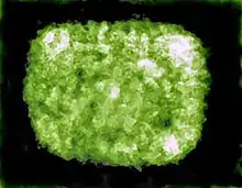
Like other viruses in the Poxviridae family, capripoxviruses are brick-shaped. Capripoxvirus virions are different than orthopoxvirus virions in that they have a more oval profile, as well as larger lateral bodies. The average size of capripoxvirions is 320 nm by 260 nm.
Genome
The virus has a 151-kbp genome, consisting of a central coding region which is bounded by identical 2.4 kbp-inverted terminal repeats and contains 156 genes.[4] There are 146 conserved genes when comparing LSDV with chordopoxviruses of other genera.[4] These genes encode proteins which are involved in transcription and mRNA biogenesis, nucleotide metabolism, DNA replication, protein processing, virion structure and assembly, and viral virulence and host range.[4] Within the central genomic region, LSDV genes share a high degree of collinearity and amino acid identity with the genes of other mammalian poxviruses.[4] Examples of viruses with similar amino acid identity include suipoxvirus, yatapoxvirus, and leporipoxvirus.[4] In terminal regions, however, collinearity is interrupted.[4] In these regions, poxvirus homologues are either absent or share a lower percentage of amino acid identity.[4] Most of these differences involve genes that are likely associated with viral virulence and host range.[4] Unique to Chordopoxviridae, LSDV contains homologues of interleukin-10 (IL-10), IL-1 binding proteins, G protein-coupled CC chemokine receptor, and epidermal growth factor-like protein, which are found in other poxvirus genera.[4]
Epidemiology
LSDV mainly affects cattle and zebus, but has also been seen in giraffes, water buffalo, and impalas.[5] Fine-skinned Bos taurus cattle breeds such as Holstein-Friesian and Jersey are the most susceptible to the disease. Thick-skinned Bos indicus breeds including the Afrikaner and Afrikaner cross-breeds show less severe signs of the disease.[3] This is probably due to the decreased susceptibility to ectoparasites that Bos indicus breeds exhibit relative to Bos taurus breeds.[6] Young calves and cows at peak lactation show more severe clinical symptoms, but all age-groups are susceptible to the disease.[3]
Transmission
Outbreaks of LSDV are associated with high temperature and high humidity [7] It is usually more prevalent during the wet summer and autumn months, especially in low-lying areas or near bodies of water, however, outbreaks can also occur during the dry season.[3] Blood-feeding insects such as mosquitos and flies act as mechanical vectors to spread the disease. A single species vector has not been identified. Instead, the virus has been isolated from Stomoxys, Biomyia fasciata, Tabanidae, Glossina, and Culicoides species.[3] The particular role of each of these insects in the transmission of LSDV continues to be evaluated.[3] Outbreaks of lumpy skin disease tend to be sporadic since they are dependent upon animal movements, immune status and wind and rainfall patterns, which affect the vector populations.[2]
The virus can be transmitted through blood, nasal discharge, lacrimal secretions, semen and saliva. The disease can also be transmitted through infected milk to suckling calves.[3] In experimentally infected cattle, LSDV was found in saliva 11 days after the development of fever, in semen after 22 days, and in skin nodules after 33 days. The virus is not found in urine or stool. Like other pox viruses, which are known to be highly resistant, LSDV can remain viable in infected tissue for more than 120 days.
Immunity
Artificial immunity
There have been two different approaches to immunization against LSDV. In South Africa, the Neethling strain of the virus was first attenuated by 20 passages on the chorio-allantoic membranes of hens' eggs. Now the vaccine virus is propagated in cell culture. In Kenya, the vaccine produced from sheep or goatpox viruses has been shown to provide immunity in cattle.[3] However, the level of attenuation required for safe use in sheep and goats is not sufficient for cattle. For this reason the sheeppox and goatpox vaccines are restricted to countries where sheeppox or goatpox is already endemic since the live vaccines could provide a source of infection for the susceptible sheep and goat populations.
In order to ensure adequate protection against LSDV, susceptible adult cattle should be vaccinated annually. Approximately, 50% of cattle develop swelling (10–20 millimetres (1⁄2–3⁄4 in) in diameter) at the site of inoculation.[3] This swelling disappears within a few weeks. Upon inoculation, dairy cows may also exhibit a temporary decrease in milk production.[3]
Natural immunity
Most cattle develop lifelong immunity after recovery from a natural infection.[3] Additionally, calves of immune cows acquire maternal antibody and are resistant to clinical disease until about 6 months of age.[3] To avoid interference with maternal antibodies, calves under 6 months of age whose dams were naturally infected or vaccinated should not vaccinated. On the other hand, calves born from susceptible cows are also susceptible and should be vaccinated.
History
Lumpy skin disease was first seen as an epidemic in Zambia in 1929. Initially, it was thought to be the result of either poisoning or a hypersensitivity to insect bites. Additional cases occurred between 1943 and 1945 in Botswana, Zimbabwe, and the Republic of South Africa. Approximately, 8 million cattle were affected in a panzootic infection in South Africa in 1949, causing enormous economic losses. LSD spread throughout Africa between the 1950s and 1980s, affecting cattle in Kenya, Sudan, Tanzania, Somalia, and Cameroon.
In 1989 there was an LSD outbreak in Israel. This outbreak was the first instance of LSD north of the Sahara desert and outside of the African continent.[2] This particular outbreak was thought to be the result of infected Stomoxys calcitrans being carried on wind from Ismailiya in Egypt. During a period of 37 days between August and September 1989, fourteen of the seventeen dairy herds in Peduyim became infected with LSD.[7] All of the cattle as well as small flocks of sheep and goats in the village were slaughtered.[7]
Throughout the past decade, LSD occurrences have been reported in Middle Eastern, European, and west Asian regions.[2]
LSD was first reported to the Bangladesh livestock department in July 2019.[8] Eventually 500,000 head were estimated to have been infected in this outbreak.[8] The Food and Agriculture Organization (FAO) recommended mass vaccination.[8] As a result of the introduction of fall armyworm and this cattle plague within a few months of each other, the FAO, the World Food Programme, Bangladesh Government officials, and others agreed to begin improving Bangladesh's livestock disease surveillance and emergency response capabilities.[8] Method of entry of the virus into Bangladesh remains unknown.[9]
In 2022 a lumpy skin disease outbreak in Pakistan killed over 7000 cattle.[10] In India between July-September 2022 the lumpy skin disease outbreak in India resulted in the death of over 80,000 cattle.[11][12] The state of Rajasthan has seen a majority of the deaths.[13] Inter-state and inter-district movement of cattle in a number of states has been restricted.[14][15][16] Indian Council of Agricultural Research labs have undertaken creation of an indigenous vaccine.[17] A goat pox vaccine is being used, 15 million doses had been administered by September 2022.[18] There are at least three centres manufacturing the goat pox vaccine in India.[19][20][21] Institutions with authority to test have been expanded.[22]
References
- Şevik, Murat; Avci, Oğuzhan; Doğan, Müge; İnce, Ömer Barış (2016). "Serum Biochemistry of Lumpy Skin Disease Virus-Infected Cattle". BioMed Research International. 2016: 6257984. doi:10.1155/2016/6257984. ISSN 2314-6133. PMC 4880690. PMID 27294125.
- http://www.oie.int/fileadmin/Home/eng/Health_standards/tahm/2.04.13_LSD.pdf
- Coetzer, J.A.W. (2004). Infectious Diseases of Livestock. Cape Town: Oxford University Press. pp. 1268–1276.
- Tulman, E. R.; Afonso, C. L.; Lu, Z.; Zsak, L.; Kutish, G. F.; Rock, D. L. (1 August 2001). "Genome of Lumpy Skin Disease Virus". Journal of Virology. 75 (15): 7122–7130. doi:10.1128/JVI.75.15.7122-7130.2001. ISSN 0022-538X. PMC 114441. PMID 11435593.
- Carter, G.R.; Wise, D.J. (2006). "Poxviridae". A Concise Review of Veterinary Virology. Retrieved 25 July 2006.
- Ibelli, A. M. G.; Ribeiro, A. R. B.; Giglioti, R.; Regitano, L. C. A.; Alencar, M. M.; Chagas, A. C. S.; Paço, A. L.; Oliveira, H. N.; Duarte, J. M. S. (25 May 2012). "Resistance of cattle of various genetic groups to the tick Rhipicephalus microplus and the relationship with coat traits". Veterinary Parasitology. 186 (3): 425–430. doi:10.1016/j.vetpar.2011.11.019. hdl:11449/4968. PMID 22115946.
- Yeruham, I; Nir, O; Braverman, Y; Davidson, M; Grinstein, H; Haymovitch, M; Zamir, O (22 July 1995). "Spread of Lumpy Skin Disease in Israeli Dairy Herds". The Veterinary Record. 137–4 (4): 91–93. doi:10.1136/vr.137.4.91. PMID 8533249. S2CID 23409535.
- "Co-ordinating a response to Fall Armyworm and Lumpy Skin Disease in Bangladesh, FAO in Bangladesh". Food and Agriculture Organization of the United Nations. 23 January 2020. Retrieved 12 February 2021.
- Badhy, Shukes Chandra; Chowdhury, Mohammad Golam Azam; Settypalli, Tirumala Bharani Kumar; Cattoli, Giovanni; Lamien, Charles Euloge; Fakir, Mohammad Aflak Uddin; Akter, Shamima; Osmani, Mozaffar Goni; Talukdar, Faisol; Begum, Noorjahan; Khan, Izhar Ahmed; Rashid, Md Bazlur; Sadekuzzaman, Mohammad (2021). "Molecular characterization of lumpy skin disease virus (LSDV) emerged in Bangladesh reveals unique genetic features compared to contemporary field strains". BMC Veterinary Research. 61 (2021). 17 (1): 61. doi:10.1186/s12917-021-02751-x. ISSN 1746-6148. PMC 7844896. PMID 33514360.
How such a virus has emerged suddenly in Bangladesh remains unknown.
- Hanif, Haseeb (9 September 2022). "Lumpy skin disease kills 7,500 cattle". The Express Tribune. Retrieved 3 October 2022.
- Bajeli-Datt, Kavita (23 September 2022). "Current outbreak of lumpy skin disease distinct from 2019, need large-scale genomic surveillance: Study". The New Indian Express. Retrieved 24 September 2022.
- Jolly, Bani; Scaria, Vinod (24 September 2022). "The evolution of lumpy skin disease virus". The Hindu. ISSN 0971-751X. Retrieved 24 September 2022.
- Rao, Lingamgunta Nirmitha (21 September 2022). Goswami, Sohini (ed.). "Lumpy skin disease: Lakhs of cattle suffer, Rajasthan worst-hit". Hindustan Times. Retrieved 3 October 2022.
- "Lumpy skin disease: Haryana bans interstate, interdistrict movement of cattle". Hindustan Times. 21 August 2022. Retrieved 24 September 2022.
- Bhusari, Piyush (9 September 2022). "Lumpy Skin Disease: Maha Now Bans Interstate Cattle Transport & Markets". The Times of India. Retrieved 24 September 2022.
- "UP Government Bans Cattle Trade With 4 States to Prevent Lumpy Skin Disease". TheQuint. PTI. 23 September 2022. Retrieved 24 September 2022.
{{cite web}}: CS1 maint: others (link) - Shagun (8 September 2022). "Lumpy skin disease outbreak: Indigenous vaccine still awaits emergency-use clearance". Down to Earth. Retrieved 24 September 2022.
- "Over 67,000 cattle died so far from lumpy skin disease in India: Centre". Business Standard. Press Trust of India. 12 September 2022. Retrieved 24 September 2022.
{{cite web}}: CS1 maint: others (link) - "Hester Bio spurts on ensuring adequate supply of Goat Pox Vaccine in India". Business Standard India. Capital Market. 26 September 2022. Retrieved 3 October 2022.
{{cite news}}: CS1 maint: others (link) - "Indian Immunologicals launches Goat Pox Vaccine". Business Line. The Hindu. 19 December 2021. Retrieved 3 October 2022.
- Keval, Varun (15 September 2022). "Telangana only state manufacturing goat pox vaccine in India". Telangana Today. Retrieved 24 September 2022.
- "Farm varsity lab gets nod for Lumpy Skin Disease testing". Hindustan Times. 24 September 2022. Retrieved 24 September 2022.
External links
- Current status of Lumpy skin disease worldwide at OIE. WAHID Interface - OIE World Animal Health Information Database
- Disease card
- Lumpy Skin Disease Food and Agriculture Organization of the United Nations