Kidneys: The Main Osmoregulatory Organ
The kidneys are a pair of bean-shaped structures that are located just below and posterior to the liver in the peritoneal cavity . Adrenal glands, also called suprarenal glands, sit on top of each kidney. Kidneys regulate the osmotic pressure of a mammal's blood through extensive filtration and purification in a process known as osmoregulation. All the blood in the human body is filtered many times a day by the kidneys. These organs use almost 25 percent of the oxygen absorbed through the lungs to perform this function. Oxygen allows the kidney cells to efficiently manufacture chemical energy in the form of ATP through aerobic respiration. Kidneys eliminate wastes from the body; urine is the filtrate that exits the kidneys.
Kidneys' location and function
Kidneys filter the blood, producing urine that is stored in the bladder prior to elimination through the urethra. They are located in the peritoneal cavity.
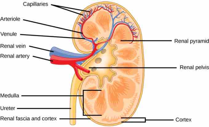
Externally, the kidneys are surrounded by three layers . The outermost layer, the renal fascia, is a tough connective tissue layer. The second layer, the perirenal fat capsule, helps anchor the kidneys in place. The third and innermost layer is the renal capsule. Internally, the kidney has three regions: an outer cortex, a medulla in the middle, and the renal pelvis in the region called the hilum of the kidney. The hilum is the concave part of the bean-shape where blood vessels and nerves enter and exit the kidney; it is also the point of exit for the ureters.

Structure of the kidney
Externally, the kidney is surrounded by the renal fascia, the perirenal fat capsule, and the renal capsule. Internally, the kidney is most importantly filled with nephrons that filter blood and generate urine.
Because the kidney filters blood, its network of blood vessels is an important component of its structure and function. The arteries, veins, and nerves that supply the kidney enter and exit at the renal hilum. Renal blood supply starts with the branching of the aorta into the renal arteries (which are each named based on the region of the kidney they pass through) and ends with the exiting of the renal veins to join the inferior vena cava. The renal arteries split into several segmental arteries upon entering the kidneys. Each segmental artery splits further into several interlobar arteries that enter the renal columns, which supply the renal lobes. The interlobar arteries split at the junction of the renal cortex and medulla to form the arcuate arteries. The arcuate, "bow shaped" arteries form arcs along the base of the medullary pyramids. Cortical radiate arteries, as the name suggests, radiate out from the arcuate arteries, branch into numerous afferent arterioles, and then enter the capillaries supplying the nephrons.