Electric Activity in the Heart
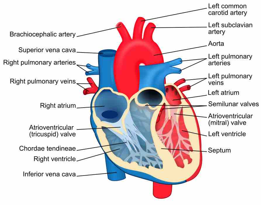
The human heart provides continuous blood circulation through the cardiac cycle and is unsurprisingly one of the most vital organs in the human body. The heart is divided into four main chambers: the two upper chambers are called the left and right atria (singular atrium) and two lower chambers are called the right and left ventricles. As can be seen in , there is a thick wall of muscle separating the right side and the left side of the heart called the septum. Normally with each beat the right ventricle pumps the same amount of blood into the lungs that the left ventricle pumps out into the body. Physicians commonly refer to the right atrium and right ventricle together as the right heart and to the left atrium and left ventricle as the left heart.

Structure of the heart
Structure diagram of a coronal section of the human heart from an anterior view. The two larger chambers are the ventricles.
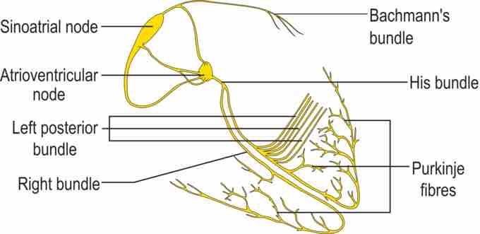
The electric energy that stimulates the heart occurs in the sinoatrial node, which produces a definite potential and then discharges, sending an impulse across the atria. In the atria the electrical signal moves from cell to cell (see section on nerve conduction and the electrocardiogram) while in the ventricles the signal is carried by specialized tissue called the Purkinje fibers which then transmit the electric charge to the myocardium. shows the isolated heart conduction system.

Conduction System of the Heart
Isolated heart conduction system, showing the sinoatrial and Purkinje fibers.
The Sinotrial Node's Role as a Pacemaker
The sinoatrial node (also commonly spelled sinuatrial node) is the impulse-generating (pacemaker) tissue located in the right atrium of the heart: i.e., generator of normal sinus rhythm. It is a group of cells positioned on the wall of the right atrium. These cells are specialized cardiomycetes (cardiac muscle cells).
The sinoatrial node (also commonly spelled sinuatrial node, abbreviated SA node) is the impulse-generating (pacemaker) tissue located in the right atrium of the heart, and thus the generator of normal sinus rhythm. It is a group of cells positioned on the wall of the right atrium. Although all of the heart's cells have the ability to generate the electrical impulses (or action potentials) that trigger cardiac contraction, the sinoatrial node normally initiates it, simply because it generates impulses slightly faster than the other areas with pacemaker potential .

Sinoatrial Node Tissue
High magnification micrograph of sinoatrial node tissue and an adjacent nerve fiber.
Cells in the SA node, located in the upper right corner of the heart, will typically discharge (create action potentials) at about 60-100 beats/minute. Because the sinoatrial node is responsible for the rest of the heart's electrical activity, it is sometimes called the primary pacemaker. If the SA node does not function, or the impulse generated in the SA node is blocked before it travels down the electrical conduction system, a group of cells further down the heart will become the heart's pacemaker. These cells form the atrioventricular node (AV node), which is an area between the atria and ventricles, within the atrial septum. If the AV node also fails, Purkinje fibers are capable of acting as the pacemaker. The reason Purkinje cells do not normally control the heart rate is that they generate action potentials at a lower frequency than the AV or SA nodes.
Purkinje Fibers
The Purkinje fibers are located in the inner ventricular walls of the heart. These fibers consist of specialized cardiomyocytes that are able to conduct cardiac action potentials more quickly and efficiently than any other cells in the heart. Purkinje fibers allow the heart's conduction system to create synchronized contractions of its ventricles, and are therefore essential for maintaining a consistent heart rhythm.
During the ventricular contraction portion of the cardiac cycle, the Purkinje fibers carry the contraction impulse from both the left and right bundle branch to the myocardium of the ventricles. This causes the muscle tissue of the ventricles to contract, thus enabling a force to eject blood out of the heart. Atrial and ventricular discharge through the Purkinje trees is assigned on a standard Electrocardiogram as the P Wave and QRS complex, respectively.
Purkinje fibers also have the ability of automatically firing at a rate of 15-40 beats per minute if left to their own devices. In contrast, the SA node outside of parasympathetic control can fire a rate of almost 100 beats per minute. In short, they generate action potentials, but at a slower rate than sinoatrial node and other atrial ectopic pacemakers. Thus they serve as the last resort when other pacemakers fail.