Concept
Version 14
Created by Boundless
Pericardium

Membranes of the Thoracic Cavity
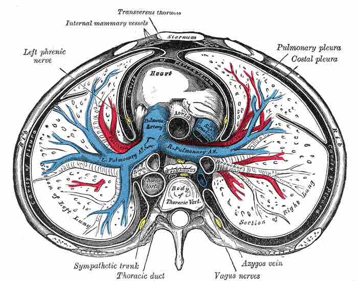
A transverse section of the thorax, showing the contents of the middle and the posterior mediastinum. The pleural and pericardial cavities are exaggerated since normally there is no space between parietal and visceral pleura and between pericardium and heart.
This transverse diagram of the thorax indicates the transverse thoracis, internal mammary vessels, left phrenic nerve, heart, rib, pleural cavity, pulmonary arteries, sympathetic trunk, thoracic duct, vagus nerves, azygos vein, pulmonary pleura, costal pleura, lungs, sternum, and thoracic aorta.
Source
Boundless vets and curates high-quality, openly licensed content from around the Internet. This particular resource used the following sources:
"Transverse Thorax."
http://upload.wikimedia.org/wikipedia/commons/0/01/Gray968.png
Wikimedia
Public domain.