White matter is one of the two components of the central nervous system. It consists mostly of glial cells and myelinated axons that transmit signals from one region of the cerebrum to another and between the cerebrum and lower brain centers. White matter tissue of the freshly cut brain appears pinkish white to the naked eye because myelin is composed largely of lipid tissue veined with capillaries.
White matter, long thought to be passive tissue, actively affects how the brain learns and dysfunctions. While grey matter is primarily associated with processing and cognition, white matter modulates the distribution of action potentials, acting as a relay and coordinating communication between different brain regions.
White matter is composed of bundles of myelinated nerve cell processes (or axons), which connect various grey matter areas (the locations of nerve cell bodies) of the brain to each other and carry nerve impulses between neurons. Myelin acts as an insulator, increasing the speed of transmission of all nerve signals.
Cerebral and spinal white matter do not contain dendrites, which can only be found in grey matter along with neural cell bodies, and shorter axons. White matter in non-elderly adults is 1.7-3.6% blood.
White matter is the tissue through which messages pass between different areas of gray matter within the nervous system. Using a computer network as an analogy, the gray matter can be thought of as the actual computers themselves, whereas the white matter represents the network cables connecting the computers together. The white matter is white because of the fatty substance (myelin) that surrounds the nerve fibers (axons). This myelin is found in almost all long nerve fibers and acts as an electrical insulation. This is important because it allows the messages to pass quickly from place to place.
There are three different kinds of tracts, or bundles of axons that connect one part of the brain to another and to the spinal cord, within the white matter:
- Projection tracts extend vertically between higher and lower brain and spinal cord centers. They carry information between the cerebrum and the rest of the body. The cortico spinal tracts, for example, carry motor signals from the cerebrum to the brainstem and spinal cord.
- Commissural tracts cross from one cerebral hemisphere to the other through bridges called commissures. Commissural tracts enable the left and right sides of the cerebrum to communicate with each other.
- Association tracts connect different regions within the same hemisphere of the brain. Among their roles, association tracts link perceptual and memory centers of the brain.
The brain in general (and especially a child's brain) can adapt to white-matter damage by finding alternative routes that bypass the damaged white-matter areas, and can therefore maintain good connections between the various areas of gray matter.
White matter forms the bulk of the deep parts of the brain and the superficial parts of the spinal cord . Aggregates of gray matter such as the basal ganglia and brain stem nuclei are spread within the cerebral white matter. The cerebellum is structured in a similar manner as the cerebrum, with a superficial mantle of cerebellar cortex, deep cerebellar white matter (called the "arbor vitae") and aggregates of grey matter surrounded by deep cerebellar white matter (dentate nucleus, globose nucleus, emboliform nucleus, and fastigial nucleus). The fluid-filled cerebral ventricles (lateral ventricles, third ventricle, cerebral aqueduct, and fourth ventricle) are also located deep within the cerebral white matter.

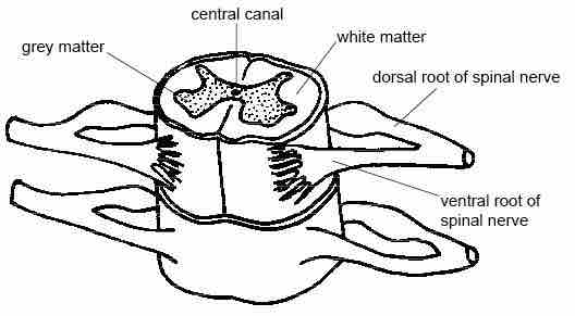
White matter in spinal cord
The spinal cord diagram showing location of the white matter surrounding grey matter.
In 1983, M. C. Raff et al. discovered that tissue samples originating from the optic nerves of rats contained two morphologically distinct types of astrocytes.
- "Type 1 astrocytes" had a fibroblasts appearance and resided in both gray matter and white matter.
- "Type 2 astrocytes" had a neuron-like appearance and resided in white matter alone.