Types of Receptors
As we exist in the world, our bodies are tasked with receiving, integrating, and interpreting environmental inputs that provide information about our internal and external environments. Our brains commonly receive sensory stimuli from our visual, auditory, olfactory, gustatory, and somatosensory systems.
Remarkably, specialized receptors have evolved to transmit sensory inputs from each of these sensory systems. Sensory receptors code four aspects of a stimulus:
- Modality (or type)
- Intensity
- Location
- Duration
Receptors are sensitive to discrete stimuli and are often classified by both the systemic function and the location of the receptor.
Sensory receptors are found throughout our bodies, and sensory receptors that share a common location often share a common function. For example, sensory receptors in the retina are almost entirely photoreceptors. Our skin includes touch and temperature receptors, and our inner ears contain sensory mechanoreceptors designed for detecting vibrations caused by sound or used to maintain balance.
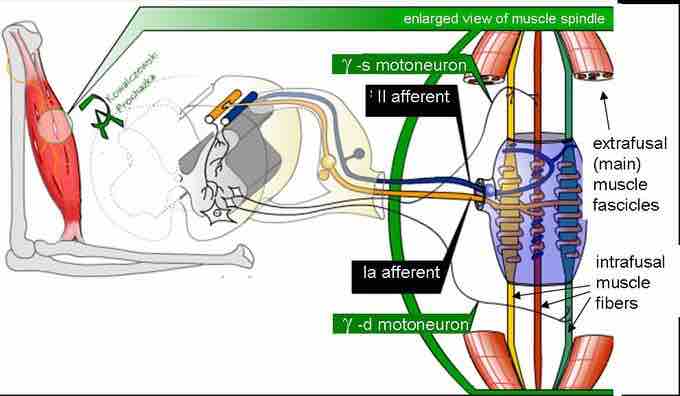
Force-sensitive mechanoreceptors provide an example of how the placement of a sensory receptor plays a role in how our brains process sensory inputs. While the cutaneous touch receptors found in the dermis and epidermis of our skin and the muscle spindles that detect stretch in skeletal muscle are both mechanoreceptors, they serve discrete functions.
In both cases, the mechanoreceptors detect physical forces that result from the movement of the local tissue, cutaneous touch receptors provide information to our brain about the external environment, while muscle spindle receptors provide information about our internal environment.

Muscle spindle
Mammalian muscle spindle showing typical position in a muscle (left), neuronal connections in spinal cord (middle), and an expanded schematic (right). The spindle is a stretch receptor with its own motor supply consisting of several intrafusal muscle fibers. The sensory endings of a primary (group Ia) afferent and a secondary (group II) afferent coil around the non-contractile central portions of the intrafusal fibers.