Fontanelles are soft spots on a baby's head that, during birth, enable the bony plates of the skull to flex and allow the child's head to pass through the birth canal. The ossification of the bones of the skull causes the fontanelles to close over a period of 18 to 24 months; they eventually form the sutures of the neurocranium.
The cranium of a newborn consists of five main bones: two frontal bones, two parietal bones, and one occipital bone. These are joined by fibrous sutures that allow movement that facilitates childbirth and brain growth.
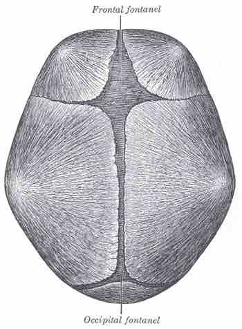
At birth, the skull features a small posterior fontanelle (an open area covered by a tough membrane) where the two parietal bones adjoin the occipital bone (at the lambda). This fontanelle usually closes during the first two to three months of an infant's life. This is called intramembranous ossification. The mesenchymal connective tissue turns into bone tissue.
The much larger, diamond-shaped anterior fontanelle—where the two frontal and two parietal bones join—generally remains open until a child is about two years old. The anterior fontanelle is useful clinically, as examination of an infant includes palpating the anterior fontanelle .

Superior view of infant skull
This image shows the location of the anterior (frontal) and posterior fontanelles.
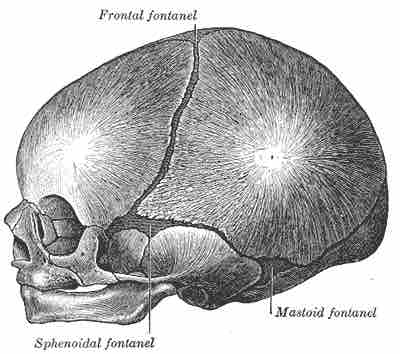
Two smaller fontanelles are located on each side of the head. The more anterior one is the sphenoidal (between the sphenoid, parietal, temporal, and frontal bones), while the more posterior one is the mastoid (between the temporal, occipital, and parietal bones) .

Lateral view of infant skull
This image show the location of the sphenoidal and mastoid fontanelles.
The fontanelle may pulsate. Although the precise cause of this is not known, it is perfectly normal and seems to echo the heartbeat, perhaps via the arterial pulse within the brain vasculature, or in the meninges. This pulsating action is how the soft spot got its name: fontanelle means little fountain.
Parents may worry that their infant may be more prone to injury at the fontanelles. In fact, although they may colloquially be called soft spots, the membrane covering the fontanelles is extremely tough and difficult to penetrate.
The fontanelles allow the infant brain to be imaged using ultrasonography. Once they are closed, most of the brain is inaccessible to ultrasound imaging because the bony skull presents an acoustic barrier.