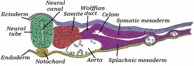
The urogenital system arises during the fourth week of development from urogenital ridges in the intermediate mesoderm on each side of the primitive aorta. The nephrogenic ridge is the part of the urogenital ridge that forms the urinary system. Three sets of kidneys develop sequentially in the embryo: The pronephros is rudimentary and nonfunctional, and regresses completely. The mesonephros is functional for only a short period of time and remains as the mesonephric (Wolffian) duct. The metanephros remains as the permanent adult kidney. It develops from the uteric bud, an outgrowth of the mesonephric duct, and the metanephric mesoderm, derived from the caudal part of the nephrogenic ridge. Urine excreted into the amniotic cavity by the fetus forms a major component of the amniotic fluid. Urine formation begins towards the end of the first trimester (weeks 11 to 12) and continues throughout fetal life. The kidneys develop in the pelvis and ascend during development to their adult anatomical location at T12-L3. This normally happens by the ninth week. The adrenal medulla forms from neural crest cells that migrate into the fetal cortex and differentiate into chromaffin cells. The urinary bladder develops from the upper end of the urogenital sinus, which is continuous with the allantois. It is lined with endoderm. The lower ends of the metanephric ducts are incorporated into the wall of the urogenital sinus and form the trigone of the bladder. The connective tissue and smooth muscle surrounding the bladder are derived from adjacent splanchnic mesoderm. The allantois degenerates and remains in the adult as a fibrous cord called the urachus (median umbilical ligament).

Intermediate mesoderm
Intermediate mesenchyme or intermediate mesoderm is a type of embryological tissue called "mesoderm" that is located between the paraxial mesoderm and the lateral plate. It develops into the part of the urogenital system (kidneys and gonads), as well as the reproductive system.