Acute Arterial Occlusion
- Article Author:
- David Smith
- Article Editor:
- Craig Lilie
- Updated:
- 8/12/2020 6:11:31 PM
- For CME on this topic:
- Acute Arterial Occlusion CME
- PubMed Link:
- Acute Arterial Occlusion
Introduction
Acute arterial occlusion is synonymous with acute limb ischemia and is considered a vascular emergency. Acute limb ischemia is defined as a sudden loss of limb perfusion for up to 2 weeks after the initiating event. Acute arterial occlusion can occur in any peripheral artery of the upper and lower extremities. Acute occlusion can lead to a limb or life-threatening ischemia. Diagnostic measures, treatment, and management depend on the artery affected and the patient's past medical history. Acute arterial occlusion is time-sensitive and left untreated can quickly progress to infarction and loss of limb and life. Acute arterial occlusion is associated with increased morbidity, significant disability, and emergent operation in high-risk patients. [1][2][3]
Etiology
The most common cause of acute limb ischemia is in situ thrombotic occlusion. It is more common in the lower extremities, and the initiating event is a preexisting history of peripheral artery disease (PAD). Thrombotic occlusions can occur in any segment of the upper and lower extremities but most commonly affects the superficial femoral artery. Patients who have a preexisting history of PAD tend to have the well-established development of collateral vessels creating variability in symptoms and severity. Other causes include embolic occlusion from the left heart, aorta, and iliac vessel, as well as penetrating or blunt trauma. [4]
Epidemiology
Published studies regarding the true incidence of acute limb ischemia are lacking. In a prospective population-based study in Oxford, England over a 10-year time frame the incidence of critical limb ischemia was determined to be 22 per 100,000 per year. Acute limb ischemia affects older men and women equally with a median age of 75 years. Risk factors include age, smoking, diabetes, obesity, sedentary lifestyle, family history of vascular disease, high cholesterol, and high blood pressure. Non-traumatic ischemia of the lower extremities is more common than the upper extremities and is more likely to result in limb loss.
Pathophysiology
In situ thrombosis most commonly affects the femoral or popliteal arteries, while emboli most commonly lodge in aortic, iliac, femoral, and popliteal bifurcations. Once arterial blood flow is impeded, under perfused tissues switch from aerobic to anaerobic metabolism, causing lactate production, acidosis, and an increased concentration of free radicals. Prolonged and untreated ischemia will cause the hypoxic muscle to deplete its ATP stores; this is followed by dysfunction in the sodium/potassium-ATPase and calcium/sodium pump leading to intracellular calcium leakage into the myocytes. Increasing free calcium negatively interacts with the muscle's myosin, actin, and proteases and eventually causes muscle fiber necrosis. Intracellular potassium, phosphate, creatinine kinase, and myoglobin are released into the systemic circulation as a result of the necrotic muscle.
History and Physical
The classic presentation of limb ischemia is known as the "six Ps," pallor, pain, paresthesia, paralysis, pulselessness, and poikilothermia. These clinical manifestations can occur anywhere distal to the occlusion. Most patients initially present with pain, pallor, pulselessness, and poikilothermia. Pain is often localized and less severe when the limb is in the dependent position. As the ischemia prolongs, paresthesia replaces pain, and the final stages of injury cause paralysis. Patients with embolic occlusion tend to have an abrupt onset with more severe symptoms, as collateralization of the vasculature has not occurred at this point. It is important to realize that symptoms can develop over the course of hours to days and present as new or recurring.
Evaluation
Initial evaluation of acute arterial occlusion begins with the physical exam. A detailed physical examination should assess for changes in temperature along the extremity affected with a bilateral comparison. A complete bilateral pulse exam should be initially conducted with manual palpation and include an arterial Doppler exam when the deficient pulse is noted. Muscle strength and sensation testing should also be assessed. When occlusion has been identified, confirmatory testing with CT angiogram (CTA) is generally conducted as it is quicker and more readily available. However, the time required to order, perform, and interpret imaging must be balanced against the urgency for treatment to preserve life and limb. Since most episodes of acute limb ischemia present to the emergency department, an immediate vascular surgery consult must be initiated. The severity of ischemia can be further assessed with the physical exam alone, utilizing the Rutherford Classification of Acute Limb Ischemia. Finding the cause of occlusion is also important. For example, a patient with a history of atrial fibrillation with abrupt initial symptoms is more likely to have an embolic occlusion as opposed to the patient with a history of PAD and claudication who is more prone to having a thrombotic source. [5]
Treatment / Management
Cases of limb-threatening ischemia require emergent vascular surgery consult. The surgical approach is directed at the reperfusion of the affected extremity. This can be accomplished by the surgical bypass, endarterectomy, or embolectomy. Results are variable and will ultimately depend on the duration of the ischemia and the extent of occlusion. Catheter-directed thrombolysis (CDT) by an interventional radiologist is becoming more common and is reserved for patients with a salvageable limb (Rutherford class II, IIa, and IIb). Patients presenting with profound paralysis and absent pain with inaudible arterial and venous pulses are considered to have irreversible damage and will require amputation. While the patient waits for surgical or interventional radiology assessment, initial therapy should include fluid resuscitation, pain control, and administration unfractionated heparin to minimize thrombus propagation. The general goal of anticoagulation is to achieve an increase in partial thromboplastin by a factor of 2.0 to 2.5 above baseline. Management after revascularization requires close and frequent observation. Prolonged ischemia of greater than 4 hours increases the risk of compartment syndrome and must be monitored with hourly neurovascular checks. [6][7][8]
Differential Diagnosis
- Arterial embolus
- Acute arterial thrombosis
- Aortic dissection
- Chronic exertional compartment syndrome
- Chronic venous insufficiency
- Degenerative disk disease
- Osteoarthritis
- Spinal stenosis
- Thromboangiitis obliterans
- Varicose vein surgery
Staging
The Society for Vascular Surgery has created a classification of an acutely ischemic limb:
- Class I: Non-threatened extremity; elective revascularization may or may not be necessary.
- Class II: Threatened extremity; immediate revascularization is indicated to prevent tissue loss.
- Class III: Ischemia has progressed to infarction, and salvage of the extremity is not possible.
Complications
- Compartment syndrome
- Amputation
- Necrosis and gangrene
- Bleeding
- Stroke
- Myocardial infarction
- Death
Postoperative and Rehabilitation Care
Once the acutely ischemic limb has been managed, these patients need to be monitored in the ICU. Aspirin or an oral anticoagulant may be required if the patient has peripheral vascular disease or atrial fibrillation. The cause of the ischemic limb must be investigated to prevent a recurrence. Since many of these patients are frail, physical therapy should be involved in the restoration of function.
Deterrence and Patient Education
- Discontinue smoking
- Control hypertension
- Manage diabetes
- Maintain a healthy weight
- Remain compliant with medications
Pearls and Other Issues
Reperfusion injury may result in the target limb after therapy. This can include profound edema, causing increased compartmental pressures. Signs and symptoms of reperfusion injury include severe pain, focal weakness, and hypoesthesia. Laboratory studies may reveal elevated creatine kinase and myoglobinuria. Most cases of compartment syndrome secondary to reperfusion injury involve the anterior compartment of the leg; therefore, assessment of peroneal nerve function is critical. Reperfusion injury also can cause cardiac, renal, and pulmonary complications making prompt recognition and treatment just as important as the initial diagnosis of acute arterial occlusion.
Enhancing Healthcare Team Outcomes
Patients with an acutely ischemic limb need an interprofessional approach with collaboration with a team consisting of a vascular surgeon, interventional radiologist, nurse, infectious disease, and critical care. Early therapy is critical if one wants to salvage the limb. In addition, the majority of these patients have other comorbidities like heart disease, obstructive lung disease, diabetes, and obesity, which also affect survival. The patient is best managed in the ICU, where the vital signs and peripheral pulses can be closely monitored by a nurse. The American College of Radiology has established guidelines that are evidence-based to determine the type of imaging in these patients.[4][9] [Level 5]
For those not deemed to be surgical candidates, catheter-directed thrombolysis may be undertaken. The pharmacist must be fully aware of all the indications and contraindications of this therapy to prevent additional morbidity. For those who undergo thrombolysis or an angiogram, the nurse is responsible for monitoring the leg for viability and bleeding in the groin, where access to the artery was obtained.
Outcomes
The outcomes for patients with an acutely ischemic limb are guarded. Even if the limb is salvaged, these patients have other comorbidities, which can lead to a heart attack, renal failure, stroke, or wound infection. Unless an interprofessional team is involved, the mortality for these individuals remains high. Expert opinion is highly recommended when these patients present to the emergency room because the earlier the comorbidity is managed, the lower the risk of complications.[10][11] [Level 5]
(Click Image to Enlarge)
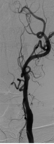
Occlusion of the internal carotid artery (ICA) results in collateral formation via the external carotid artery (ECA).
Contributed by Scott Dulebohn, MD
(Click Image to Enlarge)

ECG of Acute MI with RCA occlusion.
Contributed by Tammy J. Toney-Butler, AS, RN, CEN, TCRN, CPEN
(Click Image to Enlarge)
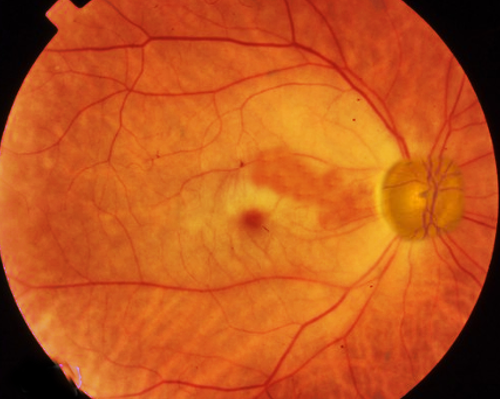
central retinal artery occlusion
Image courtesy S bhimji MD
(Click Image to Enlarge)
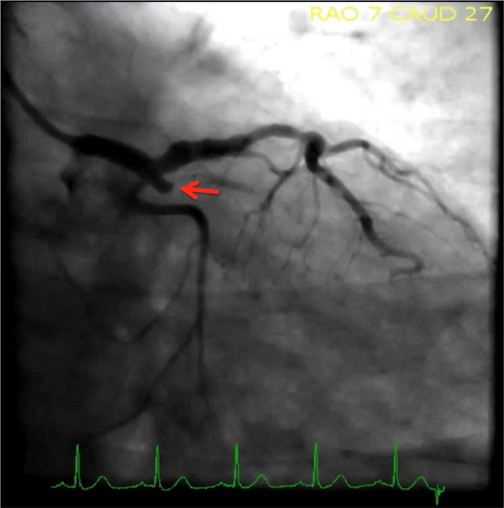
100% proximal circumflex artery occlusion, TIMI flow 0
Contributed by Alexander Bracey, MD
(Click Image to Enlarge)
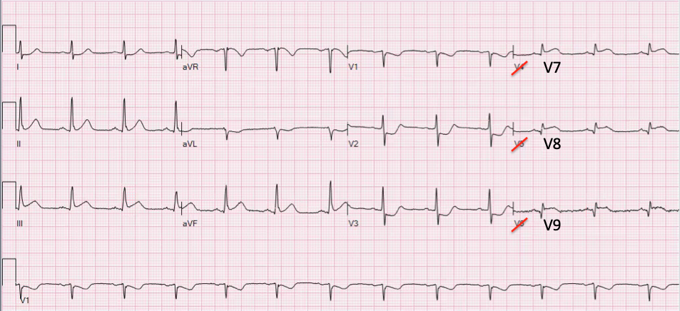
Isolated posterior MI posterior leads with STE, cath proven 100% proximal circumflex artery occlusion with TIMI 0 flow (same pt as in example 2)
Contributed by Alexander Bracey, MD