Adamantinoma
- Article Author:
- Faten Limaiem
- Article Author:
- Dawood Tafti
- Article Editor:
- Ahmad Malik
- Updated:
- 10/27/2020 9:46:18 PM
- For CME on this topic:
- Adamantinoma CME
- PubMed Link:
- Adamantinoma
Introduction
Adamantinoma is a rare low-grade malignant bone tumor of uncertain histogenesis which occurs commonly in the diaphyses and metaphyses of the tibia. The term adamantinoma has been given to this tumor due to its histological resemblance to ameloblastoma of the mandible. [1] Its histopathology shows biphasic patterns of epithelial cells and osteofibrous components. There are two types of adamantinoma: the classical and the differentiated type, which resembles osteofibrous dysplasia. [2] Despite advances in imaging techniques, the definitive diagnosis of adamantinoma is mainly established by histopathological examination.
Etiology
The origin of adamantinoma is unknown. A classically understood pathogenesis suggests early displacement of basal epithelial cells of the skin during embryological development. [3] This theory is supported by the fact that the anterior tibia, where the enchondral formed bone is closest to the skin surface, is the most common site of involvement of adamantinoma. Based on immunohistochemical and ultrastructural studies, there is a suggestion that adamantinoma may be of epithelial origin. [3] The possible relationship between adamantinoma and osteofibrous dysplasia is still a matter of debate. The potential link between these two lesions has implications for the diagnosis, prognosis, and treatment. [3][4]
Epidemiology
Adamantinoma is a rare tumor accounting for approximately 0.4% of all primary malignant bone tumors. There is a slight male predominance with a sex-ratio of 5:4. Adamantinomas frequently occur in young to middle-aged adults between the ages of 20 to 40. The pediatric and elderly populations are rarely affected. The most common sites involved by adamantinoma are the anterior metaphysis or diaphysis of the tibia. Other sites include the fibula, ulna, femur, humerus, and radius. [5][2]
Pathophysiology
Histopathology
Macroscopic findings:
Classic adamantinoma usually presents as a cortical, well-demarcated, yellowish-grey, lobulated tumor of a firm to bony consistency with peripheral sclerosis. It may be a single lesion or occasionally multifocal. Macroscopically detectable cystic spaces are commonly filled with straw-colored or blood-like fluid.
Histological findings:
Classical adamantinomas are characterized by easily recognizable epithelial and osteofibrous components that may be mixed in various proportions and differentiation patterns. The four main differentiating patterns of classic adamantinoma are:
- Basaloid
- Tubular
- Spindle cell
- Squamous
The first two patterns are most commonly encountered, but all patterns may be present. The spindle-cell component is more often observed in recurrences, lining cystic spaces, and in metastases. The osteofibrous component is composed of storiform-oriented spindle cells. Woven bone trabeculae are usually present in, or next to the center of the lesion prominently rimmed by osteoblasts, and with varying amounts of transformation to a lamellar bone at the periphery of the tumor. Foam cells or myxoid change may be present, and mast cells or multinucleated giant cells are occasionally detected. Mitotic activity is usually low. A fifth histological pattern, the so-called osteofibrous dysplasia-like variant, is characterized by a predominance of osteofibrous tissue, in which small groups of epithelial cells are only encountered by careful search or immunohistochemistry.
Extensive sampling of adamantinoma is significant especially in the differentiated form where the epithelial component is only focally encountered.
Immunohistochemical study:
The fibrous tissue is positive for vimentin. The epithelial cells show coexpression of keratin, EMA, vimentin, p63, and podoplanin. [8][9] Estrogen, progesterone, and N-cadherins are found in classic, but not in osteofibrous dysplasia-like adamantinoma. [10] In classic adamantinomas, the epithelial component is surrounded by a continuous basement membrane consisting of collagen IV, laminin and galectin 3 [10], whereas less distinct epithelial islands show multiple interruptions or no surrounding basement membrane at all. EGF/EGFR expression is restricted to the epithelial component. FGF2/FGFR1 is present in both parts, while in culture, the cells express M-CSF and RANKL, which may contribute to the osteolysis observed. [11] [12]
History and Physical
Clinically, adamantinoma often displays a protracted clinical behavior and gradually enlarges in size. The main complaint is a slow-growing swelling with or without pain. [13] Bone deformity and pathological fracture are other features which lead the patient to seek medical attention. The patient might also present with neurological deficits when the spine is involved. [3]
Evaluation
Plain radiograph:
On x-ray, the tumor is typically well-circumscribed, cortical, multi-lobulated and osteolytic. Intra-lesional opacities, septation, and peripheral sclerosis may also be seen. Multifocality within the same bone is regularly observed. The multifocal radiolucencies which are surrounded by ring-shaped densities produce the characteristic ''soap-bubble'' appearance. [14] The lesion commonly remains intracortical and extends longitudinally, but may also destroy the cortex and invade the medullary cavity or surrounding periosteum and soft tissue. This situation is usually accompanied by a lamellar or solid periosteal reaction.
Computed tomography scan:
Computed tomography scan demonstrates the soft tissue extension and cortical involvement when they exist. However, it does not depict the intraosseous extension of adamantinomas. CT scan plays a role in the routine work-up of adamantinomas and is useful in detecting pulmonary metastases. [3]
Magnetic resonance imaging:
MRI plays a crucial role in locoregional staging since it depicts distant cortical foci, intramedullary and soft tissue extension. MRI is also a useful tool for the determination of tumor-free margins as well as the strategy for reconstructive surgery. [15]
Two morphological patterns of adamantinoma are described on MRI:
- Multiple small nodules in one or more foci
- A solitary lobulated focus
Adamantinomas usually show a low signal intensity on T1-weighted images and a high signal on T2-weighted images. However, these findings are nonspecific. [3][15][3]
Treatment / Management
The most effective treatment of adamantinoma is wide excision with clear margins [16]. After en block wide resection, reconstruction of the limb can be performed with distraction allografts, osteogenesis, non-vascularized autogenous bone grafts vascularized autografts and metallic segmental replacement. [7] Amputation for adamantinoma does not improve survival rates compared to limb-preserving surgery [7]. Because of the high rate of recurrence, curettage is not recommended. [5] Radiotherapy and chemotherapy are not effective in the treatment of adamantinoma. [17]
Differential Diagnosis
Several authors have pointed to the close relationship between differentiated adamantinoma (osteofibrous dysplasia-like adamantinoma) and osteofibrous dysplasia, which can cause differential diagnostic problems due to similar histological and radiological appearance and typical location in the tibia. [18] Clinically, radiologically and histologically adamantinoma can resemble several bone tumors including bone cysts, giant cell tumor and malignant tumors (chondrosarcoma, angiosarcoma, and metastases). [3]
Prognosis
Adamantinoma is a locally aggressive neoplasm with the potential to metastasize. After inadequate surgery, recurrence of the tumor is frequent.
The risk factors for recurrent or metastatic disease are [19]:
- Symptoms of less than 5 years duration
- Male sex
- Young age less than 20 years
- Pain at presentation
- Initial treatment by curettage or resection
- Lack of squamous differentiation of the tumor
- An increase in the epithelium-to-stroma ratio
Adamantinoma metastasizes in about 15-30% of cases by a hematogenous or lymphatic route to other sites of the body, frequently to the lungs or lymph nodes, less frequently to the bones and abdominal viscera. [3]
Complications
Consultations
- Orthopedics
- Oncology
- Surgical oncology
Deterrence and Patient Education
Patients with signs and symptoms of bone pain, soft tissue swelling, or any subjective palpable mass should seek early intervention.
Enhancing Healthcare Team Outcomes
Adamantinoma is ideally managed by an interprofessional team that consists of orthopedists, radiologists, surgical oncologists, oncologists, and pathologists as well as specialty trained nurses, and pharmacists. Correlation between gross, radiographic, and microscopic features of the lesion is crucial to establish the definitive diagnosis of adamantinoma. Despite its rarity, it is essential to recognize this bone tumor since adequate treatment in early stages result in a better prognosis. Postoperatively, patients require long term follow-up due to the possibility of tumor recurrence and distant metastases. Patients and families require ongoing coordinated education by the clinician and specialty-trained oncology nurse. They should be taught to recognize the signs and symptoms of recurrence or complications. The nurse should assist in coordinating follow-up care and be available for questions. For the best outcomes, a coordination interprofessional open communication approach will lead to the best. [Level V]
(Click Image to Enlarge)

Anterior tibial sclerotic lesion representing a biopsy proven adamantinoma.
Contributed by Dr.Douglas Byerly.
(Click Image to Enlarge)

Sclerotic lesion within the anterior tibia, representative of a biopsy proven adamantinoma.
Contributed by Dr.Douglas Byerly.
(Click Image to Enlarge)

T1 low signal intramedullary lesion within the anterior tibia representative of a biopsy proven adamantinoma.
Contributed by Dr.Douglas Byerly.
(Click Image to Enlarge)
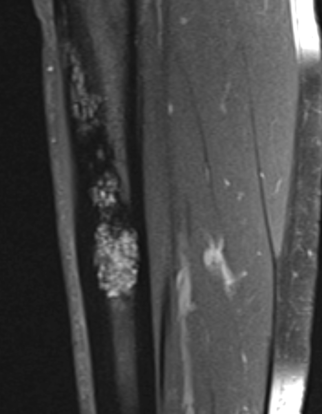
Saggital T1 with gadolinium administration. There is a cortically based lesion with heterogeneous enhancement. This was a biopsy proven adamantinoma.
Contributed by Dr.Douglas Byerly.