Anatomy, Bony Pelvis and Lower Limb, Thigh Adductor Magnus Muscles
- Article Author:
- Susan Jeno
- Article Editor:
- Gary Schindler
- Updated:
- 8/10/2020 5:46:50 PM
- For CME on this topic:
- Anatomy, Bony Pelvis and Lower Limb, Thigh Adductor Magnus Muscles CME
- PubMed Link:
- Anatomy, Bony Pelvis and Lower Limb, Thigh Adductor Magnus Muscles
Introduction
The adductor group of thigh muscles occupies the medial myofascial compartment of the thigh. This group of muscles, in general, takes origin from the pelvis and inserts on the femur. As is often the case, exceptions exist to the compartment generalizations; the gracilis, rather than attaching to the femur, attaches to the proximal medial tibia as part of the pes anserine group. The medial compartment muscles include pectineus, adductor longus, adductor brevis, gracilis and the largest of the group, adductor magnus. These muscles all serve as adductors of the thigh, but also serve as important stabilizers of the pelvis and work to maintain balance of the pelvis on the lower limb during gait. The nerve supply to most of the muscles in this compartment is the obturator nerve, which arises from the lumbar plexus from nerve roots L2-4. Adductor magnus is the largest of the medial compartment muscles; it is the most posterior of the group, and some consider it the most powerful and also the most complex of the adductor group. Adductor magnus will be the focus of this article. This muscle's complexity is in part derived from the fact that it divides into an adductor (pubofemoral) portion and a hamstring (ischiocondylar) portion.
Structure and Function
The adductor magnus is described as a large triangular muscle that has a proximal tendinous attachment to the inferior ramus of the pubis, the ramus of the ischium, and the ischial tuberosity. The part of the muscle that is considered the adductor portion has its proximal attachment on the inferior ramus of the pubis and the ramus of the ischium. The part of the muscle considered the hamstring portion has its proximal attachment on the ischial tuberosity. Each portion of the muscle has separate and distinct distal attachment points.
The adductor portion of the adductor magnus can be divided into 2 parts, a more superior part that originates on the ramus of the pubis and the lower segment which arises from the ramus of the ischium. The smaller, more horizontal portion from the pubis inserts on the medial side of the gluteal tuberosity, medial to the attachment of gluteus maximus. This portion of the muscle is in a plane which lies more anterior to the rest of the muscle, and has also been termed the adductor minimus muscle by some authors. The larger portion arises from the ramus of the ischium and extends laterally to insert into a broad aponeurosis on the linea aspera and the proximal portion of the medial supracondylar line of the femur. This larger portion of the muscle contains 5 fibrous openings that are maintained by tendinous arches. The more superior four openings, being small, allow the perforating branches and the terminal portion of the profunda femoris artery to pass into the posterior compartment of the thigh. The larger fifth opening, known as the adductor hiatus allows for the passage of the femoral vessels from the adductor canal into the popliteal fossa. The distal attachment of the adductor portion of adductor magnus on the linea aspera blends with the proximal attachment of the short head of the biceps femoris muscle. This blending creates the ability of the 2 muscles to work in a coordinated fashion as a stabilizer of the femur and pelvis. The adductor portion of the adductor magnus is innervated by the posterior division of the obturator nerve (L2,3,4).
The hamstring portion of the adductor magnus is so named due to the similarity in structure, proximal attachment, and innervation to the hamstring muscles. This portion of the adductor magnus is nearly vertical in orientation as it extends from the ischial tuberosity to the adductor tubercle on the medial femoral condyle and through fibrous attachments to the supracondylar line of the femur. This portion of the muscle is innervated by the tibial portion of the sciatic nerve (L4).
The two parts of the muscle have similar and also differing actions as both portions of the muscle are adductors of the thigh. The adductor portion of the adductor magnus, besides adduction, also flexes the thigh while the hamstring portion adducts but extends the thigh. The two portions of this muscle also function in a synergistic fashion during the gait cycle and control the pelvis for posture.
An article by Takizawa et al. examined the adductor magus of 10 limbs from embalmed cadavers, and based on the location of the perforating arteries, divided the muscle into four divisions rather than the three noted above. In their study, the authors noted that the part of the “adductor portion” that inserted on the inferior portion of the linea aspera and originated from the inferior ramus and ischial tuberosity (labeled AM3 in their study) received innervation from both the posterior division of the obturator nerve and the tibial portion of the sciatic nerve. In addition, the authors noted that the most proximal portion of the muscle has a different fiber morphology than the rest of the muscle making this uppermost segment of the muscle more suitable for stabilization activities. In contrast, the rest of the muscle with longer muscle fibers length is more functionally related to muscles such as semitendinosus and is suited for moving the lever arm of the thigh through a larger range of motion.[1] Furthermore, Takizawa et al. completed a secondary study which included 21 embalmed cadaveric limbs and found over 90% of the adductor portion (AM3) samples were supplied by both nerves. The authors also demonstrated all parts of the adductor magnus, except for the uppermost segment of the adductor portion, had dual innervation in at least some specimens. [2]
Due to the depth of the adductor portion of the adductor magnus, surface EMG studies of this large portion of the muscle (approximately 70% of the muscle) are rare. Rather, most studies of the adductor magnus focus on the hamstring portion as it is accessible for surface EMG analysis. This muscle has the potential to affect postural control of the pelvis and lower extremity during the gait cycle yet is largely unstudied.
Embryology
The embryologic development of the adductor group is first evident in the proximal region of an 11 mm embryo with separation into independent masses for the different muscles of the group including the adductor portion of the adductor magnus. When the embryo reaches 14 mm in length, the separate muscles are evident, but their tendons are not clearly separable. Concurrently, the hamstring portion of the adductor magnus differentiates from the muscle blastema of the dorsal thigh closely associated with that from which the semimembranosus muscle develops. As the embryo reaches a length of 20 mm, the muscles of the adductor compartment and those in the posterior thigh compartment along with their tendinous skeletal attachments are clearly discernable. The adductor and hamstring portions of the adductor magnus fuse at this point in development. The nerves to both portions of the adductor magnus develop from the anterior division of the lumbosacral plexus which suggests that both developmentally have a primitive flexor origin.
Blood Supply and Lymphatics
The obturator artery, a branch of the internal iliac artery, passes from within the pelvis, through the obturator foramen to enter the medial compartment of the thigh and supplies the muscles in this compartment, though the blood supply to the adductor magnus is derived from many additional vessels as well. As the perforating branches of the profunda femoris artery pass through the adductor magnus, they also are the primary source of the blood supply to the muscle. As with the other adductor muscles, the medial femoral circumflex artery supplies the superior portion of the muscle. The inferior portion of the muscle will be supplied via the femoral artery, the popliteal artery and the genicular arteries. Adductor magnus receives vascular supply from both its anterior and posterior surfaces.
Lymphatic vessels from the adductor compartment muscles, including adductor magnus, will drain into the deep inguinal lymph nodes located in the medial compartment of the femoral sheath of the femoral triangle.
Nerves
The nerves that supply the adductor magnus muscle have an embryologic origin from the anterior divisions of the lumbosacral plexus and include the obturator nerve, posterior division (L2-4), and the tibial portion of the sciatic nerve (L4). Studies by Takizawa et al. have challenged the long-held belief that the adductor portion is supplied by the posterior division of the obturator nerve, while the hamstring portion receives its supply from the tibial portion of the sciatic nerve. [1][2]
Physiologic Variants
In a 2011 study by Tubbs RS et al., the authors dissected 20 adult cadavers and five fetuses looking for the presence of adductor minimus. The study revealed that approximately half of the adult (52.5%) and all of the fetal dissected limbs demonstrated an adductor minimus muscle separate from the adductor magnus muscle and five others where the adductor minimus was partially fused to the adductor magnus muscle. This study also determined that when the adductor minimus was absent or underdeveloped, the quadratus femoris muscle extended more inferiorly, but a fascial layer maintained separation of the two muscles.[1] In an earlier study, Tubbs et al. also noted that a vastoadductor membrane, part of the intermuscular septum, can exist between the vastus medialis and the adductor magnus. The femoral artery may undergo compression by this membrane prior to the artery passing through the adductor canal.[3]
As previously indicated, a study by Takizawa et al. found the portion of the adductor portion of the muscle that inserts most distally on the femur obtains innervation from both the posterior division of the obturator nerve and the tibial portion of the sciatic nerve.[1] Other parts of the muscle may also have dual innervation.[[2]
Surgical Considerations
Obey et al.[4] studied the proximal attachments of the hamstrings and adductor magnus on the ischial tuberosity on cadaveric specimens. Avulsion injuries of the hamstrings tendon from the ischial tuberosity are increasingly recognized as an injury, in athletic populations, that does not respond well to conservative treatment, has poor outcomes, and hence are recommended for prompt surgical repair. An intact adductor magnus may mask a complete avulsion of the hamstring tendons on MRI evaluation as it appears to be an “intact but attenuated semimembranosus tendon.” The authors state an intact adductor magnus may result in a misdiagnosis of patients as having only a partial tear rather than a complete avulsion injury. They conclude that adductor magnus is located medial to the hamstring origins on the ischial tuberosity and in some specimens was a sizable structure which may lead to some of the issues associated with MRI imaging of this area. Understanding the anatomical relationships between the adductor magnus and the hamstring tendons is crucial for appropriate diagnosis and surgical vs. conservative treatment of injuries in this area. Knowledge of the adductor magnus' origin on the ischial tuberosity can aid the surgeon in the appropriate anatomical repair of avulsion injuries of the hamstring muscles. As indicated in a study by Broski et al., radiologists also need to be aware of the presence of the adductor magnus tendon on the ischial tuberosity when visualizing the ischial tuberosity on MRI to avoid confusion during diagnostic studies.[5]
Clinical Significance
A study by Arnold and Delp[6] studied the contribution of the medial hamstrings and adductor muscles in the crouched, internally rotated gait of individuals with cerebral palsy to determine how these muscles contributed to the internal rotation seen with this gait pattern. The medial hamstrings, adductor brevis/longus/hamstring portion of magnus muscles in upright standing and normal femoral anteversion were found to have a slight internal rotation moment arm, the proximal adductor magnus and gracilis also had a minor medial rotation moment arm, and the middle and distal segment of the adductor portion of magnus had a negligible rotational moment arm. The study found, however, that with hip anteversion greater than 20 degrees or knee flexion greater than approximately 30 degrees, the moment arms of these muscles changed, and most became more externally biased. This finding is an important consideration when assessing the strategies for improvement of gait in these individuals. Care must be exercised when treating muscles around the hip as positional differences can influence muscular function.
Other Issues
The adductor magnus muscle is both a dynamic stabilizer of the pelvis and femur as well as a prime mover of the femur into adduction. The adductor magnus can be likened to the deltoid muscle; one portion flexes the thigh and works as a medial rotator while the other extends the thigh and is a lateral rotator, and both portions adduct the thigh. This is similar to the anterior (flexor/medial rotator/abductor) and posterior (extensor/lateral rotator/abductor) actions of the deltoid. The curvilinear attachment of the adductor magnus on the pelvis is also reminiscent of the curvilinear attachment of the deltoid on the spine of the scapula, acromion, and clavicle. Additionally, both muscles serve as dynamic stabilizers of the ball and socket joints with which they are associated, shoulder or hip respectively. This comparison may aid students in making the correlations between the functions of the upper and lower extremities.
(Click Image to Enlarge)
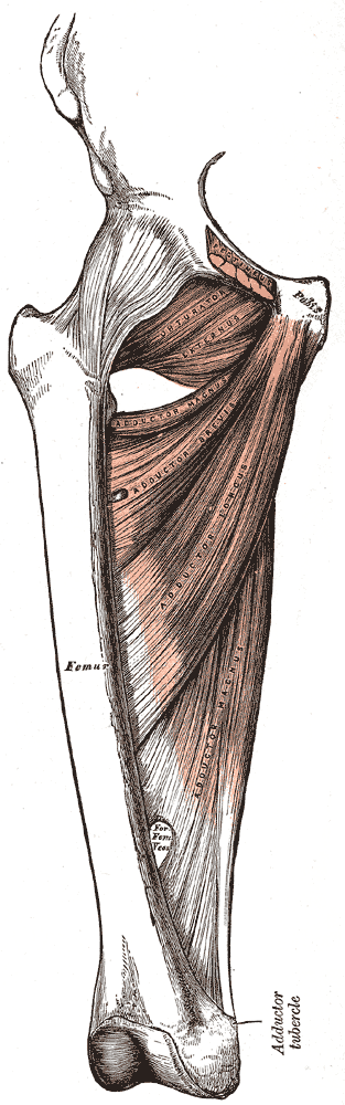
Medial Compartment of the Thigh, Pubis, Femur, Obturator Externus, Adductor Magnus; Brevis; Longus,
Contributed By Gray's Anatomy Plates