Adenoiditis
- Article Author:
- Ian Bowers
- Article Editor:
- Carl Shermetaro
- Updated:
- 8/12/2020 3:21:52 PM
- For CME on this topic:
- Adenoiditis CME
- PubMed Link:
- Adenoiditis
Introduction
The adenoids are a grouping of lymphoid tissue located on the posterior wall of the nasopharynx behind the soft palate. The adenoids, along with the faucial tonsils, lingual tonsils, and tubal tonsils of Gerlach make up what is known as Waldeyer’s ring. Together, these tissues function as an essential part of the human immune system. Antigens, introduced through the oral and nasal cavities, come into contact with the immune cells of Waldeyer's ring. These cells can then produce immunologic memory of the antigens and fight them by producing IgA antibodies; this is thought to result in a "priming" of the immune system in infancy[1].
The adenoids are present at birth and enlarge throughout childhood, reaching peak size by age seven. In most individuals, they will regress in size during puberty and may be nearly absent by adulthood. For this reason, adenoiditis is commonly a problem of childhood and adolescence. Adenoiditis occurs when there is inflammation of the adenoid tissue resulting from infection, allergies, or irritation from stomach acid as a component of LPR. Adenoiditis rarely occurs on its own and is more often involved in a more extensive disease process such as adenotonsillitis, pharyngitis, rhinosinusitis, etc. Continual irritation may lead to adenoid hypertrophy which is responsible for many of the complications of adenoid disease. Adenoiditis can be classified as acute or chronic.
Clinical Anatomy
The adenoids receive their blood supply from the ascending pharyngeal artery, maxillary artery, and facial artery. Venous drainage occurs through the pharyngeal veins. Nervous innervation is through the vagus nerve and glossopharyngeal nerve. Adenoid size grading is on a scale of zero to four[2]:
- 0 absent
- 1+ <25% obstruction of the nasopharynx
- 2+ 25-50% obstruction
- 3+ 50-75% obstruction
- 4+ >75% obstruction
Etiology
Many agents and pathogens can cause inflammation of the adenoid tissue. A viral upper respiratory tract infection (URI) often precedes acute adenoiditis. In this vulnerable state, bacterial pathogens can infect the tissues and proliferate.
The most common bacterial pathogens cultured from adenoid specimens are[3]:
- Haemophilus influenza
- Streptococcus pneumoniae
- Streptococcus pyogenes
- Staphylococcus aureus
Chronic adenoiditis is more often a polymicrobial infection and may include anaerobic pathogens and frequently results from biofilm development.[4]
Allergies are believed to play a role in adenoiditis and subsequent adenoid hypertrophy. Allergens inhaled through the nose come in contact with the adenoid tissue. The tissues will proliferate in order to create a response to allergens and produce IgA.[5]
Chronic irritation from stomach acid in the setting of gastroesophageal reflux disease (GERD) may also play a role in adenoiditis and adenoid hypertrophy, particularly in infants and young children.[6]
Epidemiology
Exact incidence and prevalence statistics for adenoiditis alone are difficult to elucidate, as adenoiditis is usually addressed in the context of a more extensive disease process such as rhinosinusitis and adenotonsillar disease. Since adenoid tissue atrophies during puberty, adenoiditis is typically a disease of children. Current literature does not suggest a predilection for gender, race, region, socioeconomic class in this disease, though parental smoking has been positively correlated[7].
Adenoiditis can be challenging to differentiate from bacterial sinusitis in children. Statistics on sinusitis in children, therefore, may give us some idea of the frequency of adenoiditis. Estimates are that children have six to eight viral URIs per year. Five to thirteen percent of these viral URIs result in bacterial superinfection, leading to sinusitis with adenoiditis as a potential component of the illness.[8]
Pathophysiology
Acute adenoiditis often occurs after a viral upper respiratory tract infection (URI). Bacterial agents proliferate and infect the adenoids and surrounding tissue resulting in inflammation and increased production of exudates. Symptoms include rhinorrhea, post-nasal drip, nasal obstruction, snoring, fever, and halitosis. Chronic adenoiditis shows many of the same symptoms but on a persistent basis lasting 90 days and is often caused by polymicrobial infections and biofilm formation. Exudates are frequently absent in chronic adenoiditis.[4]
Another cause of adenoiditis is environmental allergens or caustic irritation from stomach acid in the presence of GERD/LPR.[6]
Any form of chronic inflammation may lead to the proliferation of lymphoid tissue and subsequent adenoid hypertrophy. This hypertrophy can lead to nasal airway obstruction and obstruction of the Eustachian tubes which in turn leads to other problems such as obstructive sleep apnea (OSA) and otitis media.[3]
History and Physical
Adenoid tissue typically regresses around puberty. Therefore, the typical patient with adenoiditis is a prepubescent child with a recent history of URI. The patient may also have a history of recurrent acute otitis media, chronic nasal obstruction with mouth-breathing, chronic otitis media, sleep-disordered breathing/obstructive sleep apnea, or GERD/LPR.
Physical findings include purulent rhinorrhea, post-nasal drip, nasal obstruction, snoring, fever, mouth breathing, and halitosis. Indirect mirror exam may allow the practitioner to observe enlarged adenoids with exudates, though this can be a very challenging exam to perform in children. A flexible nasal and laryngeal endoscopic exam can allow for better evaluation of the adenoids but can require advanced training to use as well as the cooperation of the child and parents.
Long-standing adenoiditis with subsequent adenoid hypertrophy in early childhood can lead to the development of what is known as adenoid facies, or long face syndrome. Enlarged adenoids block the nasopharynx and result in obligate mouth breathing, which can lead to craniofacial abnormalities including a high-arched palate and retrognathic mandible.[9]
Evaluation
Clinical Evaluation
The diagnosis of acute adenoiditis is made clinically based on the findings of:
Possible concurrent acute otitis mediaFeverPurulent rhinorrheaPost-nasal dripNasal obstruction Throat painHalitosis
Visual inspection of the adenoids may be attempted using a laryngeal mirror or nasal endoscope.
Laboratory TestingRapid strep testCulturesAllergy testing
If it presents in the context of pharyngitis, the clinician may want to perform a rapid strep test. The purpose of doing so is two-fold. First, this will give a definitive diagnosis of the patient’s condition and help guide antibiotic therapy. Second, the doctor’s office will have a record of positive and negative strep tests which will play an important role when deciding whether an adenoidectomy, plus or minus tonsillectomy, is indicated. It is important to remember that adenoiditis remains a clinical diagnosis, so if the strep test is negative the physician can presume it is due to a different causative organism.
In cases of persistent infection despite antibiotic therapy, the clinician may choose to perform throat cultures to help identify the causative agent and guide therapy as direct cultures of adenoids may be difficult in the office setting.
If the adenoiditis is believed to be the result of seasonal or environmental allergies, allergy skin testing may be useful in directing therapy.
Radiology Testing
Lateral neck X-ray
Computed tomography (CT) of the sinuses
Sinus X-rays or sinus CTs may be obtained to look for a source of infection in the sinuses if this is suspected clinically. This is rarely required in routine cases. Lateral neck X-rays are an effective way to evaluate specifically for adenoid hypertrophy. In a patient with adenoid hypertrophy who snores a sleep study can be obtained to rule out obstructive sleep apnea.
Treatment / Management
Adenoiditis is often seen clinically as a component of rhinosinusitis or pharyngitis. Due to this fact, practitioners often use clinical management guidelines for rhinosinusitis and pharyngitis when approaching the treatment of adenoiditis.
Medical Management
Watch and waitIf the clinician believes the cause of adenoiditis is by the common cold or other common viral infection they should refrain from using antibiotics. Typically, uncomplicated upper respiratory viral infections will resolve within five to seven days.[8]
Antibiotic treatment
If symptoms continue or clinical presentation is suggestive of bacterial etiology, such as a high fever or purulent discharge from the nose or throat, the first-line management is antibiotics covering the most common pathogens. Amoxicillin is a commonly used first-line agent due to its good coverage and tolerability. Alternatively, cefdinir or cefuroxime may be used, particularly if the patient has not responded to amoxicillin. If the patient has a penicillin allergy, alternatives include clarithromycin or azithromycin. Effective antibiotic treatment should yield an improvement of symptoms in 48-72 hours. Treatment duration should be ten days, as treating for a shorter duration yields significant relapse rates and breeds antibiotic resistance. If the condition fails to improve after a course of amoxicillin or other first-line agents, amoxicillin-clavulanate should be prescribed to eliminate potential beta-lactamase producing organisms.[8]
Allergy treatment
If the adenoiditis is believed to be secondary to environmental allergies, the patient can be given a trial of nasal steroid sprays, oral steroids, oral antihistamines, or some combination thereof to see if this produces any relief in symptoms. If this is effective, the patient may benefit from formal allergy testing followed by immune-modulating therapy to provide definitive relief.
Reflux treatment
If the adenoiditis is believed to be secondary to LPR/GERD, treatment of this condition using lifestyle and diet modification with or without the use of H2 blockers or proton-pump inhibitors may provide sufficient relief of symptoms.[6]
Surgical Management
Adenoidectomy
In the absence of symptomatic improvement after treatment with amoxicillin-clavulanate or if the patient has multiple episodes of adenoiditis requiring antibiotic treatment, referral to an otolaryngologist is warranted for further evaluation and potential surgical intervention. Depending on the individual circumstances, surgical procedures may include adenoidectomy with or without tonsillectomy or myringotomy with tympanostomy tube placement, or endoscopic sinus surgery. If the patient meets the Paradise criteria for tonsillectomy, most otolaryngologists will remove the adenoids at the same time to remove another possible source of recurrent infections.[10]
Differential Diagnosis
Differential diagnosis includes:
- Viral URTI
- Sinusitis
- Rhinosinusitis
- Nasal polyposis
- Pharyngitis
- Tonsillitis
- Seasonal/environmental Allergies
- Nasopharyngeal neoplasm
- Laryngopharyngeal reflux
Prognosis
The medical treatment available for treating adenoiditis is successful in most instances. For those with recurrent disease, adenoidectomy provides a definitive solution by removing the hypertrophic or infected adenoid tissue.
Complications
If adenoiditis is left untreated, the patient may develop a chronic infection of the adenoids which in some cases can lead to the development of a biofilm. The adenoids may then serve as a nidus of infection for other closely related structures and lead to rhinosinusitis, pharyngitis, tonsillitis, and otitis media.[4][11]
Adenoid Hypertrophy
Adenoid hypertrophy is responsible for some of the more common complications related to disease of the adenoids. As they enlarge the tissues can create a significant obstacle to the flow of air through the nasopharynx. This enlargement can cause mouth breathing, snoring, and OSA. OSA can be a life-threatening disease if left untreated. Removing the adenoids can increase the flow of air through the nasopharynx, decreasing obstructive episodes, and leading to better CPAP compliance or resolution of the condition altogether.
Enlarged adenoids may also obstruct the opening of the Eustachian tubes in the nasopharynx. Without proper function of the Eustachian tube, negative pressure can build in the middle ear. This negative pressure can lead to the formation of an effusion which can cause conductive hearing loss and speech problems, as well as serve as a nidus for bacterial infections.
Long-standing adenoiditis with subsequent adenoid hypertrophy can lead to the development of what is known as adenoid facies or long-face syndrome. Enlarged adenoids can block the nasopharynx and result in obligate mouth breathing, which can lead to craniofacial abnormalities including a high-arched palate and retrognathic mandible.[9]
Consultations
Patients with recurrent adenoiditis or the complications of adenoid hypertrophy should obtain a referral to an otolaryngologist for further evaluation and treatment. Other disciplines that may need to be involved in the care of the patient include sleep medicine, allergy specialists, and gastroenterology depending on the individual’s needs.
Deterrence and Patient Education
Adenoiditis is a common issue in children and may be unavoidable since they are frequently in contact with the common pathogens and allergens that cause the inflammation. However, it is essential to seek treatment before chronic adenoiditis, and adenoid hypertrophy develop, as these can lead to serious complications and decreased quality of life.
Pearls and Other Issues
- Adenoiditis is a condition of childhood as most adenoid tissue atrophies by adulthood.
- Adenoiditis is seldom a solitary issue. It is usually discussed as being part of or indistinguishable from adenotonsillitis, rhinosinusitis, or pharyngitis.
- Adenoid hypertrophy is responsible for the most common health issues associated with the adenoids.
- Obstructive sleep apnea (OSA) is the most serious complication of adenoid disease.
- Adenoidectomy is the definitive treatment for adenoid disease.
Enhancing Healthcare Team Outcomes
Because causes of adenoiditis can include a number of different factors including recurrent bacterial infections, allergies, and GERD, treatment of adenoiditis and its complications may require the care of multiple specialists. These specialists should work in close coordination to maximize patient outcomes. It is important to find the root cause/s and treat them, or the problem may never fully resolve and lead to further complications. Healthcare team members should pay close attention to the signs and symptoms of OSA as this is the most serious complication of adenoid disease.
(Click Image to Enlarge)
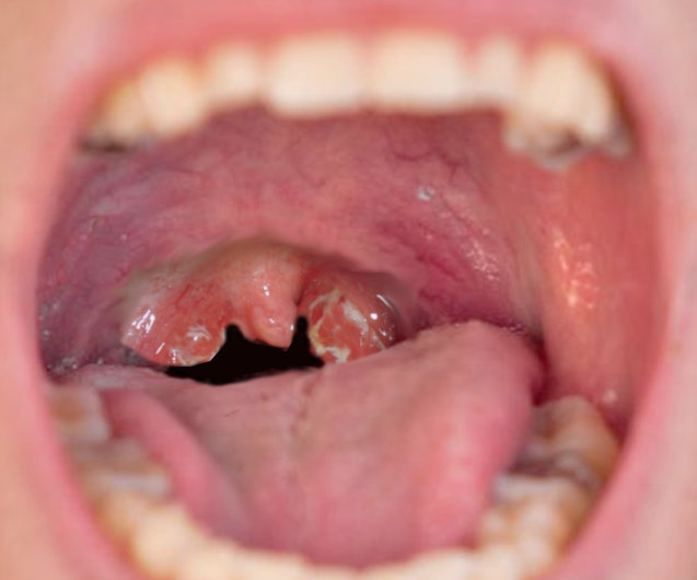
Adenoiditis
Image courtesy S Bhimji MD