Alopecia
- Article Author:
- Ahmad Al Aboud
- Article Editor:
- Patrick Zito
- Updated:
- 9/29/2020 4:09:09 PM
- For CME on this topic:
- Alopecia CME
- PubMed Link:
- Alopecia
Introduction
The term alopecia means hair loss regardless of the cause. It is not exclusive to the scalp; it can be anywhere on the body. Everyone is born with hundreds of thousands of hair on the head. The hair cycle consists of three phases: the growth phase, which is called anagen, the resting phase, which is called catagen, and the shedding phase, which is the telogen phase.[1] Ninety percent of hair are in the growth phase (anagen) and the rest, which corresponds to ten percent in the resting and shedding phases.[2] When hair is falling out, this is the telogen phase, and the hair is going to recycle, and it starts growing again in the (growth) anagen phase.[3] Alopecia can be subdivided into two main categories: scarring and non-scarring.
The most common type is non-scarring or androgenetic alopecia. The majority of men start to lose hair in the twenties, while women begin to lose their hair in their forties or fifties. As an individual grows older, they will lose hair. The difference between male hair loss and female hair loss is the pattern. Men generally lose hair in the front and the temporal region, while women tend to lose hair from the central area of the scalp. Also, female hair loss will not end up with complete baldness, whereas male hair loss can end up with complete baldness. Males tend to retain hair at the posterior area of the scalp because this hair is resistant to the androgenic hormone.[4]
Etiology
Non-scarring alopecia falls into six major categories:
1) Alopecia areata: This is hair loss that can affect every part of the body, including the scalp, face, trunk, and extremities. When it affects only a portion of the body, it is called alopecia areata. When it affects an entire site, it is called alopecia totalis. When it involves the whole body, it is called alopecia universalis. The etiology is unknown, but it might be related to an autoimmune disease.[5]
2) Androgenetic alopecia: is a pattern of hair loss that is affected by the genes and hormones (androgenic).
3) Telogen effluvium: results from shifting of the hair cycle growth (anagen) phase towards the shedding (telogen) phase. It may result from an illness like hypo or hyperthyroidism. Also, it can arise from stress like major surgery. A crash diet, poor feeding, and drugs can cause telogen effluvium.[6]
4) Traumatic alopecia: This is similar to traction alopecia, which results from forceful traction of the hair commonly seen in children. Also, trichotillomania is a type of traumatic alopecia in which the patient pulls on his/her hair repeatedly.[7]
5) Tinea capitis: the classical kind of tinea capitis (black-dots) causes non-scarring hair loss, unlike other types like kerion and favus.
6) Anagen effluvium: This is hair shedding that occurs during the anagen phase of the cell cycle. Seen in cancer patients who are receiving chemotherapeutic agents.
Scarring alopecia is divided into three major types:
1) Tinea capitis: the inflammatory variety of tinea capitis (favus) may culminate with scarring alopecia.
2) Alopecia mucinosa: This occurs when mucinous material accumulates in the hair follicles and the sebaceous glands. The mucinous material causes an inflammatory response that hinders the growth of hair.
3) Alopecia neoplastica: This is the metastatic infiltration of the scalp hair with malignant cells.
Epidemiology
The epidemiology is variable depending on the cause of alopecia and the type. In alopecia areata, the prevalence is 0.2% with no racial or sexual predilection, and it may affect any age group. Androgenetic alopecia is a common disorder affecting 50% of men and 15% of women, especially postmenopausal women.[8] It affects white races more than dark races. In telogen effluvium, women tend to be affected more than men. In tinea capitis, the incidence is much higher in the pediatric age group and is more common in dark-skin individuals with no sexual predilection.[9] Anagen effluvium is common in cancer patients treated with chemotherapeutic agents.
Pathophysiology
The pathophysiology is dependent on the type of alopecia. In alopecia areata, it is unknown, but the most common hypothesis involves autoimmunity in the form of a T-cell–mediated pathway. In androgenetic alopecia, both genetic and hormonal androgens play a role in the pathogenesis. In telogen effluvium, the shedding of hair is under the influence of hormone or stress, but sometimes the trigger is not very clear.[10] The dermatophyte infection is responsible for hair loss in tinea capitis. In anagen effluvium, the shedding of hair is under the effect of chemotherapeutic agents. In alopecia mucinosa, the infiltration of the scalp with abnormal lymphocytes is the cause.[11]
Histopathology
In patients with alopecia areata, there is a peribulbar lymphocytic infiltrate with a decrease in the ratio of anagen to telogen hair. In androgenetic alopecia, there are miniaturized hair follicles with an increase in the telogen-to-anagen ratio without inflammatory reaction. Telogen effluvium is characterized by an increase in the number of catagen hair. In tinea capitis, there is evidence of fungal infection as under a microscope along with a neutrophilic infiltrate. In anagen effluvium, there is a decrease in anagen hair without any inflammatory response. Finally, in alopecia mucinosa, there is an infiltrate of the epidermis, dermis, and peribulbar lymphocytic infiltrate mainly anaplastic cells.[12]
History and Physical
In a patient with alopecia, an accurate history helps in determining the diagnosis. The clinician should ask about the number of hair loss per day and the onset of the issue. In cases of alopecia areata, hair loss can happen overnight, while some other types of alopecia may require months or years to become apparent. Also, the clinician should ask about the general health of the patient, including his/her diet, habits, and overall general health, including recent labs, especially iron, thyroid, ovarian, and male hormones. The patient bathing habits should be asked, including shampooing and the use of conditioner. Stress is a significant cause of hair loss, and the clinician should inquire about it as well. Family history may provide a clue for patients who may have a genetic predisposition to hair loss (androgenetic alopecia).
During the physical examination, it is essential to notice the pattern of hair loss. In a patient with androgenetic alopecia, patients tend to lose hair from the frontal and temporal area (male type) and the central scalp area (female type). In alopecia areata, the patient may lose hair from a single area (alopecia areata classical type), the whole scalp and eyebrows (alopecia totalis), or from the entire body (alopecia universalis). In tinea capitis, the classic presentation is black dots associated with broken hair, while the inflammatory type (favus) correlates with the scarring type of alopecia. Telogen effluvium classically presents with diffuse thinning of hair, and the pull test will be positive. In the pull test, ten hair are gently pulled from ten different spots, and if three or more hair from these spots are removed, then it would be positive and indicate the underlying cause of hair loss; if less than three hair are removed, then the test is negative. In a patient with alopecia mucinosa, the patient would have multiple flesh-colored papules and nodules infiltrating the skin of the scalp.
Evaluation
Patients with alopecia need to be investigated thoroughly to determine the type and cause. Complete blood count, iron panel, thyroid function test, autoantibodies, total testosterone, and free testosterone, ovarian hormones, luteinizing hormone, and the follicular stimulating hormone may be necessary for some patients. KOH preparation and fungal culture in case of tinea capitis are mandatory. Chest x-ray and MRI are required in the case of alopecia mucinosa to stage the disease (mycosis fungoides).
Evaluation of the hair pulled, and hair loss can reveal information such as if the strands are broken, whether the bulb of the hair is white or dark (indicating telogen cycle vs. anagen cycle). A hair pull test is a simple investigation performed in the office. Clinicians may also use dermoscopy/trichoscopy. A biopsy will reveal the most information regarding hair loss.
Hair Pull Test
- Test for excessive shedding, it is not diagnostic of a particular hair loss but indicates active hair loss is occurring.
- It is performed by gently grasping 40-60 hairs and gently pull upward from different parts of the scalp.
- A positive test is six or more strands, although some clinicians use three.
- The pull test is positive in:
- telogen effluvium
- anagen effluvium
- androgenetic alopecia
- alopecia areata and scarring alopecia
- loose anagen syndrome
Treatment / Management
The provider should direct the treatment plans according to the etiology and type of alopecia. In androgenetic alopecia, anti-androgen medications like finasteride along with minoxidil spray will help, but ultimately the patient might require a hair transplant.[13] In alopecia areata, medium potency corticosteroids, along with minoxidil spray and topical immunomodulators like tacrolimus, may benefit the patient. In tinea capitis, the mainstay of treatment is antifungal medications.
Differential Diagnosis
Non-scarring Alopecia
- Androgenetic alopecia
- Alopecia areata
- Telogen effluvium
- Traction alopecia
- Trichotillomania
- Anagen effluvium
Scarring Alopecia
- Alopecia mucinosa
- Metastatic infiltrate
- Favus
Prognosis
According to studies, around 8.5% of patients with alopecia totalis and universalis achieved a complete recovery. Many of the patients will obtain at least a transient recovery of partial or total hair regrowth. Response to treatment is often unpredictable, and healthcare providers should be aware of the prognosis and its effects to properly counsel patients.[14]
The Alopecia Areata Predictive Score is a new trichoscopy-based assessment to predict the treatment outcome in patients with patchy alopecia areata.[15]
Complications
Patients with alopecia are at increased risk of psychosocial complications of hair loss such as anxiety and depression. At the same time, these patients need to be assessed for other autoimmune conditions such as thyroid conditions, vitiligo, etc.
One study found that patients with alopecia areata are at increased risk of developing insulin resistance. [16]
Enhancing Healthcare Team Outcomes
Healthcare providers should evaluate all cases of alopecia by obtaining a thorough history and conducting a complete physical exam. Patients should be instructed not to wash their hair 24 to 48 hours before the visit to ensure an accurate pull test. The hair should be examined under a Woods light to check for any elements of fungal infection. If there is any uncertainty about the diagnosis or management, the patient should be referred to a dermatologist. The interprofessional team can optimize the treatment of these patients through communication and coordination of care. Primary care physicians, dermatologists, and nurse practitioners provide diagnoses and care plans. Specialty care nurses and pharmacists should work with the team to provide patient education. Pharmacists should evaluate medications prescribed, recognize drug-drug interactions, and monitor compliance reporting concerns to the team. The interprofessional team can thus improve outcomes for patients with alopecia. [Level V]
(Click Image to Enlarge)

Alopecia Areata
Contributed by DermNetNZ
(Click Image to Enlarge)
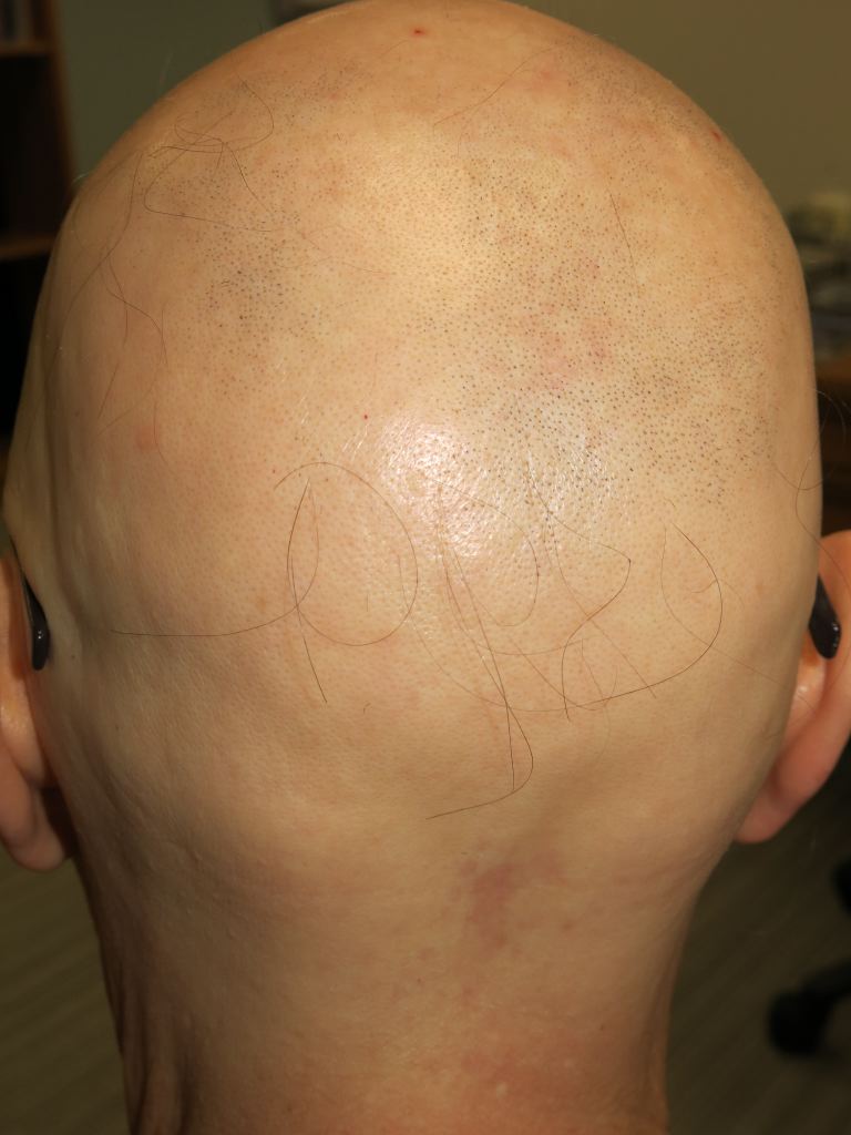
Alopecia Totalis
Contributed by DermNetNZ
(Click Image to Enlarge)
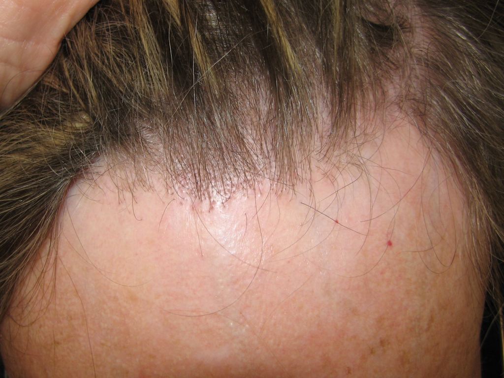
Frontal Fibrosing Alopecia
Contributed by DermNetNZ
(Click Image to Enlarge)
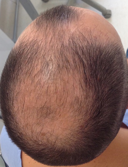
male-pattern androgenetic alopecia with loss of hair from frontal, temporal and central scalp areas.
Contributed by Ahmad Al Aboud, MD
(Click Image to Enlarge)
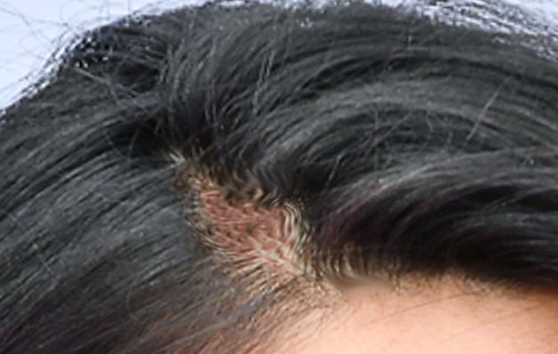
Traction alopecia
Image courtesy S Bhimji MD