Apocrine Hidrocystoma
- Article Author:
- Wissem Hafsi
- Article Author:
- Talel Badri
- Article Editor:
- Farhan Shah
- Updated:
- 9/9/2020 6:03:04 PM
- For CME on this topic:
- Apocrine Hidrocystoma CME
- PubMed Link:
- Apocrine Hidrocystoma
Introduction
Apocrine hidrocystoma is a rare, benign, cystic tumor of the apocrine sweat glands. It most commonly presents as a solitary, asymptomatic, papule or nodule and is most often located on the head and neck. Occasionally, it may present in other areas. Multiple forms are rare and may be an important marker for some rare inherited diseases. Practitioners make the diagnosis of apocrine hidrocystoma histologically, and surgical removal is the most common treatment for cystic lesions.[1][2][3][4][5]
Etiology
The etiology of apocrine hidrocystoma is unknown.
Epidemiology
Apocrine hidrocystoma is mainly observed in adults aged 30 to 70 years and rarely occurs during childhood or adolescence. Solitary apocrine hidrocystoma is equally noticed in males and females, and there is no ethnic predominance or geographic region predilection. Multiple forms of apocrine hidrocystoma are rare in the general population. This condition is not associated with a familial incidence.
Pathophysiology
The pathogenesis of apocrine hidrocystoma is not entirely known. The tumor derives from the secretory part of apocrine sweat glands. Apocrine hidrocystoma is currently considered to be a cystic proliferation of apocrine glands, rather than a simple retention cyst.[6][7][8]
Multiple apocrine hidrocystomas have been described in two rare ectodermal dysplasias: a particular form of Goltz-Gorlin syndrome and Schopf-Schultz-Passarge syndrome. Goltz-Gorlin syndrome (Focal dermal hypoplasia) is an X-linked dominant disease characterized by linear skin atrophy, microcephaly, microphthalmia, midfacial hypoplasia, malformation of the ears, mental retardation, and skeletal abnormalities. Some additional clinical findings were described in a particular form combining multiple apocrine hidrocystomas, bilateral keratoconus, esophageal papillomatosis and hiatus hernia. Schopf-Schultz-Passarge syndrome is an autosomal recessive condition characterized by the association of multiple eyelid apocrine hidrocystomas with hypotrichosis, hypodontia, palmar and plantar hyperkeratosis, as well as nail fragility.
History and Physical
Apocrine hidrocystoma is most often a solitary tumor, although practitioners have also documented multiple lesions. Apocrine hidrocystoma is typically intradermal, moderately firm, dome-shaped, translucent, blue, bluish-black, grayish, or purple cystic nodule. Its size ranges from 3 mm to 15 mm in diameter. Giant variants measuring up to 7 cm in diameter can rarely occur. Apocrine hidrocystoma is more frequently pigmented than eccrine hidrocystoma. Its pigmented appearance may be due to a Tyndall effect and/or to the presence of lipofuscin pigments in the cystic fluid. Apocrine hidrocystoma does not change or become symptomatic in hot weather.
Apocrine hidrocystoma is usually found on the head and neck, commonly affecting the cheek and the inner canthus of the eye. Apocrine hidrocystoma has also been reported in other sites, including the chest, shoulder, axilla, umbilicus, foreskin, penal shaft, vulva, and fingers. This is probably because apocrine glands are frequent in these locations. No cases of spontaneously involuting apocrine hidrocystomas have been reported.
The differential diagnosis of solitary apocrine hidrocystoma includes eccrine hidrocystoma, epidermoid or pilar cysts, cystic basal cell epithelioma, and melanoma.
The main differential diagnosis of the rare cases of multiples apocrine hidrocystomas is multiple eccrine hidrocystomas, which affect most often females (sex ratio 8:1) of the same age group. Eccrine hidrocystomas are smaller, with a diameter ranging from 1 mm to 6 mm. Their color is brown or bluish, clearer than that of apocrine hidrocystomas. Another important differential fact is that eccrine hidrocystomas increase in size and number in summer months or heated environment and may disappear entirely in cooler weather. The differential diagnosis for multiple apocrine hidrocystomas also includes syringomas, milia, trichoepitheliomas, and angiofibromas.
Evaluation
Dermoscopic findings show a homogeneous pale gray or bluish pattern, whitish cotton wool-like structures, linear vessels, and nonconstant focal brownish orange areas. The grayish color is probably due to the presence of sialomucin which induces an effect of diffraction as seen in Kaposi sarcoma. The whitish structures are due to a reflection of the connective tissue, and the brownish orange structures could be explicated by the presence of clear cells containing important amounts of glycogen. The dermoscopic sign of the peripheral linear vessels corresponds to the dilated vessels of the papillary dermis. The main interest in dermoscopy is to make sure the absence of dermoscopic features of a malignant tumor that may have a similar clinical presentation (mainly amelanotic melanoma and basal cell carcinoma).[9][10][11][12]
Histopathological findings
Histologically, apocrine hidrocystoma presents as an adenoma located in the dermis as cystic, often multiloculated, areas. They are lined by a double cellular base, an outer layer composed of cubic myoepithelial cells forming intracavitary papillary digitations, and an internal layer composed of secreting cylindrical cells having an eosinophilic cytoplasm with a characteristic decapitation secretory apical prominence. Periodic acid-Schiff (PAS)-positive granules are observed in the presence of lipofuscin granules, S-100 protein staining is generally negative. In approximately 50% of cases, there are areas of epithelial hyperplasia with intracystic papillary proliferation, as opposed to eccrine hidrocystoma.
Apocrine hidrocystoma may be suspected clinically, but the diagnostic confirmation is histological.
Treatment / Management
The most common treatment of apocrine hidrocystoma is surgical excision with narrow margins, because of the benign nature of the lesion. This is the only approach that allows a practitioner to carry out both the final diagnosis and the treatment of these tumors. An alternative therapy is needle puncture; however, local recurrence is frequently observed with this treatment. Cyst puncture followed by hypertonic glucose sclerotherapy was used successfully in the treatment of eyelid apocrine hidrocystoma. Trichloroacetic acid injection (followed by aspiration) after cyst puncture may also be an alternative to surgery.
Other therapeutic methods that may be used are electrodesiccation, radiofrequency ablation, and carbon dioxide laser treatment. Botulinum toxin A has been used effectively in a patient.
Differential Diagnosis
- Basal cell carcinoma
- Blue nevi
- Cutaneous melanoma
- Eccrine cystadenoma
- Follicular cyst
- Milia
- Syringoma
Pearls and Other Issues
Apocrine hidrocystoma is a benign cystic lesion that typically occurs on the face. The diagnosis may be suspected from certain clinical characteristics, but the formal diagnosis is based on histological examination. Management of apocrine hidrocystoma mainly involves surgical excision; however, other treatments such as electrodesiccation, carbon dioxide laser vaporization, botulinum toxin A, and trichloroacetic acid can be tried in multiple lesions, as in the treatment of eccrine hidrocystomas.
Lesions grow gradually and do not tend to resolve after attaining full size. They rarely recur after surgical excision. Cysts may cause esthetic burden and psychological distress in patients; however, cysts are usually asymptomatic. No malignant transformation of apocrine hidrocystoma has been reported to date.
Enhancing Healthcare Team Outcomes
Apocrine hidrocystoma is a benign cystic lesion that typically occurs on the face. It is a rare lesion that may be encountered by the nurse practitioner or the primary care provider. A referral to a dermatologist is highly recommended. The diagnosis may be suspected from certain clinical characteristics, but the formal diagnosis is based on histological examination. Management of apocrine hidrocystoma mainly involves surgical excision; however, other treatments such as electrodesiccation, carbon dioxide laser vaporization, botulinum toxin A, and trichloroacetic acid can be tried in multiple lesions, as in the treatment of eccrine hidrocystomas.
(Click Image to Enlarge)
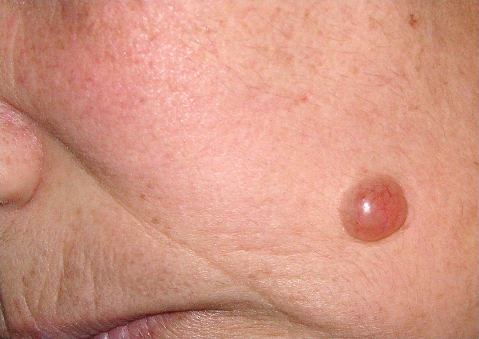
Apocrine hidrocystoma of the left cheek
Contributed by Talel Badri, MD