Appendicitis
- Article Author:
- Mark Jones
- Article Author:
- Richard Lopez
- Article Editor:
- Jeffrey Deppen
- Updated:
- 10/1/2020 11:22:41 PM
- For CME on this topic:
- Appendicitis CME
- PubMed Link:
- Appendicitis
Introduction
Appendicitis is inflammation of the vermiform appendix. This is a hollow organ located at the tip of the cecum, usually in the right lower quadrant of the abdomen. However, it can be located in almost any area of the abdomen, depending on if there were any abnormal developmental issues or if there are any other concomitant conditions such as pregnancy or prior surgeries. The appendix develops embryonically in the fifth week. During this time, there is a movement of the midgut to the external umbilical cord with the eventual return to the abdomen and rotation of the cecum. This results in the usual retrocecal location of the appendix. It is most often a disease of acute presentation, usually within 24 hours, but it can also present as a more chronic condition. If there has been a perforation with a contained abscess, then the presenting symptoms can be more indolent. The exact function of the appendix has been a debated topic. Today it is accepted that this organ may have an immunoprotective function and acts as a lymphoid organ, especially in the younger person. Other theories contend that the appendix acts as a storage vessel for "good" colonic bacteria. Still, others argue that it is a mear developmental remnant and has no real function.[1][2][3]
Etiology
The cause of appendicitis is usually from an obstruction of the appendiceal lumen. This can be from an appendicolith (stone of the appendix), or some other mechanical etiologies. Appendiceal tumors such as carcinoid tumors, intestinal parasites, and hypertrophied lymphatic tissue are all known causes of appendiceal obstruction and appendicitis. Often, the exact etiology of acute appendicitis is unknown. When the appendiceal lumen gets obstructed, bacteria will build up in the appendix and cause acute inflammation with perforation and abscess formation. One of the most popular misconceptions is the story of the death of Harry Houdini. After being unexpectedly punched in the abdomen, the rumor goes, his appendix ruptures, causing immediate sepsis and death. The facts are that Houdini did die from sepsis and peritonitis from a ruptured appendix, but it had no connection to him being struck in the abdomen. It was more related to widespread peritonitis and the limited availability of effective antibiotics at the time.[4][5]
Epidemiology
Appendicitis occurs most often between the ages of 5 and 45 with a mean age of 28. The incidence is approximately 233/100,000 people. Males have a slightly higher predisposition of developing acute appendicitis compared to females, with a lifetime incidence of 8.6% for men and 6.7 % for women. There are approximately 300,000 hospital visits yearly in the United States for appendicitis-related issues.
Pathophysiology
The pathophysiology of appendicitis likely stems from obstruction of the appendiceal orifice. This results in inflammation, localized ischemia, perforation, and the development of a contained abscess or frank perforation with resultant peritonitis. This obstruction may be caused by lymphoid hyperplasia, infections (parasitic), fecaliths, or benign or malignant tumors. When an obstruction is the cause of appendicitis, it leads to an increase in intraluminal and intramural pressure, resulting in small vessel occlusion and lymphatic stasis. Once obstructed, the appendix fills with mucus and becomes distended, and as lymphatic and vascular compromise advances, the wall of the appendix becomes ischemic and necrotic. Bacterial overgrowth then occurs in the obstructed appendix, with aerobic organisms predominating in early appendicitis and mixed aerobes and anaerobes later in the course. Common organisms include Escherichia coli, Peptostreptococcus, Bacteroides, and Pseudomonas. Once significant inflammation and necrosis occur, the appendix is at risk of perforation, leading to a localized abscess and sometimes frank peritonitis.[6]
The most common position of the appendix is retrocecal. While the anatomical position of the root of the appendix is mostly constant, tail positions can vary. Possible positions include retrocecal, subcecal, pre- and post-ileal, and pelvic.
Histopathology
Microscopic findings in acute appendicitis include the proliferation of neutrophils of the muscularis propria. The degree and extent of inflammation are directly proportionate to the severity of the infection and duration of the disease. As this condition progresses, extra appendiceal fat and surrounding tissues become involved in the inflammatory process. In severe situations, the cecum may be involved and may require resection at the time of surgery.
History and Physical
Classically, appendicitis presents as an initial generalized or periumbilical abdominal pain that then localizes to the right lower quadrant. Initially, as the visceral afferent nerve fibers at T8 through T10 are stimulated, and this leads to vague centralized pain. As the appendix becomes more inflamed, and the adjacent parietal peritoneum is irritated, the pain becomes more localized to the right lower quadrant. Pain may or may not be accompanied by any of the following symptoms:
- Anorexia
- Nausea/vomiting
- Fever (40% of patients)
- Diarrhea
- Generalize malaise
- Urinary frequency or urgency
Uncommon presentation
Some patients may present with uncommon features. In these patients, the pain may have woken the patient up from sleep. In addition, the rare patient may complain of pain while walking or coughing.
Pain upon passive extension of the right leg with the patient in the left lateral decubitus position is known as psoas sign. This maneuver stretches the psoas major muscle, which can be irritated by an inflamed retrocecal appendix. Patients often flex the hip to shorten the psoas major muscle and relieve pain.
Physical exam findings are often subtle, especially in early appendicitis.
As inflammation progresses, signs of peritoneal inflammation develop. Signs include:
- Right lower quadrant guarding and rebound tenderness over McBurney's point (1.5 to 2 inches from the anterior superior iliac spine (ASIS) on a straight line from the ASIS to the umbilicus)
- Rovsing's sign (right lower quadrant pain elicited by palpation of the left lower quadrant)
- Dunphy's sign (increased abdominal pain with coughing)
Other associated signs such as psoas sign (pain on external rotation or passive extension of the right hip suggesting retrocecal appendicitis) or obturator sign (pain on internal rotation of the right hip suggesting pelvic appendicitis) are rare.
The time course of symptoms is variable but typically progresses from early appendicitis at 12 to 24 hours to perforation at greater than 48 hours. Seventy-five percent of patients present within 24 hours of the onset of symptoms.
The risk of rupture is variable but is about 2% at 36 hours and increases about 5% every 12 hours after that.
Evaluation
The emergency department physician must refrain from giving the patient any pain medication until the surgeon has seen the patient. The analgesics can mask the peritoneal signs and lead to a delay in diagnosis or even a ruptured appendix.
Lab Testing
Elevated white blood cells (WBC) with or without a left shift or bandemia is classically present, but up to one-third of patients with acute appendicitis will present with a normal WBC count. There are usually ketones found in the urine, and the C-reactive protein may be elevated.
Imaging
Appendicitis is traditionally a clinical diagnosis. However, CT scan has greater than 95% accuracy for the diagnosis of appendicitis and is used with increasing frequency.[7][8][9]
CT criteria for appendicitis include an enlarged appendix (greater than 6 mm in diameter), appendiceal wall thickening (greater than 2 mm), peri-appendiceal fat stranding, appendiceal wall enhancement, the presence of appendicolith (approximately 25% of patients). It is unusual to see air or contrast in the lumen with appendicitis due to luminal distention and possible blockage in most cases of appendicitis. Nonvisualization of the appendix does not rule out appendicitis.
Ultrasound is less sensitive and specific than CT but may be useful to avoid ionizing radiation in children and pregnant women. MRI may also be useful for the pregnant patient with suspected appendicitis and an indeterminate ultrasound.
Classically the best way to diagnose acute appendicitis is with a good history and detailed physical exam performed by an experienced surgeon; however, it is very easy to get a CT scan done in the emergency department. It has become common practice to rely mostly on the CT report to make the diagnosis of acute appendicitis. Occasionally appendicoliths are incidentally found on routine x-rays or CT scans. These patients are at a higher risk of developing appendicitis than the general population.
These patients should be considered for prophylactic appendectomies. Studies have also shown a 10% to 30% incidence of appendicoliths present in appendectomy specimens done for acute appendicitis.
Treatment / Management
While in the emergency department, the patient must be kept NPO and hydrated intravenously with crystalloid. Antibiotics should be administered intravenously as per the surgeon. The responsibility for the consent falls on the surgeon.
The gold-standard treatment for acute appendicitis is to perform an appendectomy. Laparoscopic appendectomy is preferred over the open approach. Most uncomplicated appendectomies are performed laparoscopically. In cases where there is an abscess or advanced infection, the open approach may be needed. The laparoscopic approach affords less pain, quicker recovery, and the ability to explore most of the abdomen through small incisions. Situations, where there is a known abscess from a perforated appendix, may require a percutaneous drainage procedure usually done by an interventional radiologist. This stabilizes the patient and allows the inflammation to subside over time, enabling a less difficult laparoscopic appendectomy to be performed at a later date. Practitioners also start patients on broad-spectrum antibiotics. There is some disagreement regarding preoperative antibiotic administration for uncomplicated appendicitis. Some surgeons feel routine antibiotics in these cases are not warranted, while others give them routinely. There have also been several studies promoting the treatment of uncomplicated appendicitis solely with antibiotics and avoiding surgery altogether.[1][10]
In patients with an appendiceal abscess, some surgeons continue antibiotics for several weeks and then perform an elective appendectomy. When the appendix has ruptured, the procedure can still be done laparoscopically, but extensive irrigation of the abdomen and pelvis is necessary. In addition, the trocar sites may have to be left open.
Differential Diagnosis
The differential diagnosis includes Crohn ileitis, mesenteric adenitis, mittelschmerz, salpingitis, ruptured ovarian cyst, ectopic pregnancy, tubal-ovarian abscess, musculoskeletal disorders, endometriosis, pelvic inflammatory disease, gastroenteritis, right-sided colitis, renal colic, kidney stones, irritable bowel disease, testicular torsion, ovarian torsion, round ligament syndrome, epididymitis, and other nondescript gastroenterological issues.
Prognosis
If diagnosed and treated early, within 24 to 48 hours, the recovery and prognosis should be very good. Cases that present with advanced abscesses, sepsis, and peritonitis may have a more prolonged and complicated course, possibly requiring additional surgery or other interventions.
Complications
Postoperative abscesses, hematomas, and wound complications are all complications that can be seen after appendectomies. If the wound does get infected, one may grow Bacteroides. "Recurrent" appendicitis can occur if too much of the appendiceal stump is left after an appendectomy. This acts just like an appendix and can become occluded and infected just as with the initial episode. Therefore, it is important to ensure that there be very minimal and preferably no residual appendiceal stump after an appendectomy. If left untreated, appendicitis can lead to abscess formation with the development of an enterocutaneous fistula. Diffuse peritonitis and sepsis can also develop, which may progress to significant morbidity and possible death.
Pearls and Other Issues
Special consideration should be given to the treatment of patients with perforated appendicitis with an abscess. Those who present with an abscess and do not exhibit peritonitis may benefit from CT or ultrasound-guided percutaneous drain placement as well as antibiotics. Interval appendectomy is classically performed 6 to 10 weeks after recovery. Historically, 20% to 40 % of patients treated medically for perforated appendicitis with an abscess had recurrent appendicitis in historical literature. More recent studies suggest these rates be much lower.
Complications of appendicitis and appendectomy include surgical site infections, intra-abdominal abscess formation (3% to 4% in open appendectomy and 9% to 24% in laparoscopic appendectomy), prolonged ileus, enterocutaneous fistula, and small bowel obstruction.
Occasionally the incorrect diagnosis of acute appendicitis is made when, in reality, the correct diagnosis is Crohn disease of the cecum or terminal ileum. It is important to know that is this occurs that the appendix should be left in place if there is involvement at its base. The removal of the appendix in this situation has a high leak and fistula rate formation. On the other hand, if the base of the appendix is spared, then the appendix should be removed, even if it appears normal. This eliminates the future confusion of diagnosing acute Crohn disease versus acute appendicitis.
In the past, it was commonplace to routinely remove the appendix at the time of other nonrelated surgeries to avoid developing appendicitis in the future. Today, however, most surgeons do not routinely remove a normal appendix at the time of other scheduled procedures. If a patient does go into surgery for an incorrect diagnosis of acute appendicitis, then it is advised to remove the appendix to avoid any future diagnostic issues.
Enhancing Healthcare Team Outcomes
Patients with appendicitis usually first present to the emergency department with abdominal pain. The triage nurse should be familiar with the signs and symptoms of appendicitis because these patients need urgent admission and treatment to prevent perforation. However, making a diagnosis of appendicitis is not always easy.
Several guidelines exist that can help healthcare workers make a diagnosis of appendicitis. While most physicians, nurse practitioners, and physician assistants rely on the physical exam, others may obtain an ultrasound. For questionable cases, a CT scan of the abdomen may be helpful. The American College of Radiology recommends an ultrasound in pregnant women and an MRI in inconclusive cases in the same patient population.[11][12]
While the patient is undergoing investigation, the nurse should start an IV, administer fluids as ordered. In women, a pregnancy test must be done to rule out an ectopic. The surgeon should be notified. Pain medications should typically only be administered after the surgeon has seen the patient. The nurse should monitor the patient for acute changes in pain or vital signs and report to the interprofessional team. Before surgery, the pharmacist should evaluate for potential drug-drug interactions and potential drug allergies, reporting to the team any potential concerns.
Controversy also exists on how to best manage an appendiceal mass or phlegmon and when to undertake surgery. There is no longer any question that laparoscopic appendectomy is associated with minimal pain and faster recovery, but it is costly. Other studies indicate that a single small incision provides comparable results to a laparoscopic appendectomy and is cost-effective. Given these controversies, an interprofessional team approach to diagnosis and management of appendicitis needs to be established in each institution to ensure that the patient has no morbidity, and the management is cost-effective. [13](Level 3)
Outcomes
Many large series show that simple appendicitis treated either with an open or laparoscopic procedure has excellent outcomes. (Level 3) However, more severe and complicated appendicitis is known to be associated with worse outcomes and greater utilization of resources. Further, the atypical presentation of appendicitis in pregnancy and the elderly may also make diagnosis difficult and lead to a higher incidence of complications. [14][15](Level 3) In an era of managed care where quality care indices are monitored, it behooves healthcare workers to know the current standards of diagnosis and management of appendicitis or face denial of reimbursement.
(Click Image to Enlarge)
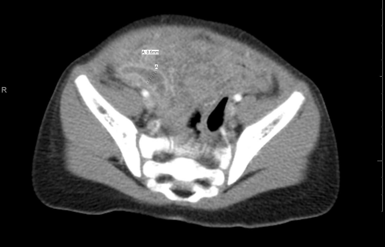
CT Abdomen Acute Appendicitis
Contributed by Scott Dulebohn, MD
(Click Image to Enlarge)

Ultrasound of the right lower quadrant with findings of acute appendicitis. There is a blind ending tubular structure measuring up to 7 mm in diameter.
Contributed by Kevin Carter, DO
(Click Image to Enlarge)

There is acute appendicitis with a dilated fluid filled tubular structure in the right lower quadrant on this axial and sagittal images with a surrounding fluid collection and stranding due to developing abscess.
Contributed by Kevin Carter, DO