Biceps Tendon Rupture
- Article Author:
- David Hsu
- Article Author:
- Prashanth Anand
- Article Editor:
- Ke-Vin Chang
- Updated:
- 11/2/2020 2:43:25 PM
- For CME on this topic:
- Biceps Tendon Rupture CME
- PubMed Link:
- Biceps Tendon Rupture
Introduction
The biceps tendon consists of 2 heads, one originating from the coracoid process (short head) and the other from supraglenoid tubercle of the scapula and superior labrum (long head). The tendon attaches to the bicipital tuberosity of the radius. The biceps tendon is a strong supinator of the forearm and serves as a weak elbow flexor. Some reports also mention the long head biceps tendon’s contribution to the stability of the glenohumeral joint. The majority of biceps rupture involves the long head.
Rupture of the proximal biceps tendon can be treated conservatively, while injury to its distal attachment usually needs surgical intervention. Patients generally recover successfully if they receive a timely diagnosis and treatment.[1][2][3]
Etiology
Distal biceps rupture is from excessive eccentric force as the arm is brought into extension from flexion. These activities include weightlifting, wrestling and labor intensive job. Proximal biceps rupture is generally not due to a unique mechanism of injury but highly correlated with rotator cuff disease.[4]
Risk factors include age, smoking, obesity, use of corticosteroids, and overuse. Rare causes include the use of quinolones, diabetes, lupus, and chronic kidney disease.
Epidemiology
The incidence of distal biceps tendon rupture is around 2.55 per 100,000 patient-years. Most patients (more than 95%) are males, and injury events usually happen during middle age(35y-54y).[5] Rupture of the distal biceps mainly involves the dominant limb. However proximal biceps rupture is commonly seen in elderly patients and its exact incidence is not known but is more common than distal biceps rupture.
Pathophysiology
Age, overuse, smoking, and corticosteroid contributes to tendon degeneration and later, tendinopathy. Furthermore, there is a vascular watershed zone at the distal biceps tendon, and lack of sufficient blood supply also plays a crucial role in potentiating tendon rupture.[6]
The proximal tendon rupture in most cases occurs at the tendon-labral junction or the bony attachment. Distal tendon ruptures usually occur at the insertion on the radial tuberosity.
Histopathology
The histopathology studies show that the torn biceps tendon exhibit an increase in proteoglycan, collagen type III, matrix metallopeptidase-1, and matrix metallopeptidase-3, disorganized fiber arrangements, which were compatible with the finding of tendinopathy.
History and Physical
Patients suffering from biceps tendon rupture may complain of sudden sharp pain in the elbow or shoulder depending on the site of rupture. Usually, there is a history of sudden eccentric force to a flexed elbow. They may feel an audible "pop" in the affected arm at the time of the injury. Pain can persist for weeks to months. Pain may diminish if the tendon is completely torn. Patients usually complain of bulbous mass in the upper arm due to excessive retraction of the biceps muscle belly and this is known as "Popeye deformity". In obese individuals, it is generally hard to appreciate this popeye deformity.
In patients with distal biceps rupture, ecchymosis, swelling, and tenderness may be present in the antecubital fossa. If the bicipital aponeurosis (lacertus fibrosus) is involved, the muscle will be retracted to the upper arm and a defect of the distal tendon will be palpated. The hook test can be used to identify the absence of the biceps tendon at its distal insertion. First, the examiner positions the patient's arm in 90 degrees of flexion and then supinates it. Second, the examiner tries to hook the tendon underneath the skin. Intact distal biceps tendon permits the examiner to hook index finger under the biceps tendon. This test has a high specificity and sensitivity in diagnosing distal biceps tear.[7]
In patients with proximal biceps tendon rupture, surprisingly there could be very little signs and symptoms other than pain. They may have ecchymosis of the proximal arm, sometimes extending up to the elbow. Proximal biceps rupture does not result in any long term change in elbow or shoulder strength. It is important to check for atrophy of shoulder girdle muscle and shoulder impingement because the proximal biceps tendon disorders are usually associated with rotator cuff pathology.
Evaluation
Diagnosis is often clinically made, while imaging is helpful when the diagnosis is unclear or partial rupture is considered. Three criteria described for diagnosis:
- History of a single traumatic event
- Grossly palpable and visible signs of retraction of the biceps muscle belly(popeye deformity)
- Weakness of flexion of the elbow and supination of the forearm in cases of distal biceps rupture
Partial ruptures may present with similar, but subtle, symptoms and physical presentation is usually with pain, weakness and no palpable defect, sometimes leading to delayed diagnosis. Ultrasound is an inexpensive, noninvasive tool to reveal the absence of the tendon but it is highly technician dependent. Radiographs generally cannot aid in diagnosis; however, it is helpful to survey for other accompanying conditions, confirm the absence of another bony pathology, or sometimes reveal radial tuberosity hypertrophy or occasional avulsion fracture of the tuberosity. MRI is rarely necessary for diagnosis, but it is helpful to distinguish between the following:
- Complete versus partial tear
- Muscle substance versus tendon tear
- Degree of retraction
Treatment / Management
Management of biceps rupture depends on the site of rupture.
Rupture of the Proximal Biceps Tendon (Long Head)
Non-surgical treatment is usually sufficient for proximal tendon rupture as it is more common in elderly patients. However, residual cosmetic deformity and some intermittent biceps muscle cramping may persist. Younger patients and female patients who are unwilling to accept cosmetic deformity and athletic patients with frequent cramping may opt for surgical intervention in the form of biceps tenodesis. The presence of associated rotator cuff pathology can also influence surgical management. Subpectoral tenodesis is the preferred approach for biceps tenodesis where the tendon is attached to the bone in the bicipital groove. Sometimes this procedure is done concurrently with arthroscopic rotator cuff pathology treatment. Newer implants like interference screws and bio-absorbable suture anchors can be placed either through the open or arthroscopic approach to secure the tendon in the subpectoral space. All the approaches as mentioned above are reported to achieve good clinical outcomes. However, until now, there is limited data to show the superiority of the surgical intervention to the non-surgical approach.[8]
Rupture of the Distal Biceps Tendon
Distal biceps rupture is usually seen in younger active patients and surgery is generally indicated for faster recovery and to return to sports.[9][10][11]. Most surgeons recommend operative treatment for rupture of the distal biceps tendon to regain the maximal strength of forearm supination and to effectively relieve pain in the antecubital fossa. Patients with low physical demands and multiple comorbidities are more suitable for conservative treatments. If the bicipital aponeurosis is intact, the functional deficits due to biceps rupture can be minimized.
Surgical repair of the distal biceps tendon can be divided into 2 methods. The non-anatomic approach indicates sutures of the ruptured biceps tendon to the brachialis, which is a simple and efficient way to regain flexion strength. The anatomic approach indicates reinsertion of the ruptured tendon on the radial tuberosity, which is reported to have better effects of restoring the strength of elbow flexion and forearm supination.
There are 2 techniques for surgical exploration and fixation of the torn distal biceps tendon.[12]
Anterior Single-Incision Technique
- Incision is over the anterior aspect of the elbow, at or just distal to antecubital fossa
- The most common complication is an injury to the lateral antebrachial cutaneous nerve. Sometimes the posterior interosseous nerve might be damaged.[13]
- Less common heterotopic ossification and synostosis compared to dual incision technique.
Dual-Incision Technique
- The method is developed to avoid injury to the posterior interosseous nerve.
- It includes a smaller anterior incision over the antecubital fossa and a second posterolateral elbow incision
- The dual-incision technique is more common to develop synostosis and heterotopic ossification than the single incision approach.
Differential Diagnosis
The diagnosis of the biceps tendon rupture sometimes is challenging. The investigator should bear in mind that proximal biceps tendon injury usually coexists with rotator cuff disorders and shoulder girdle instability.
Differential diagnosis includes:
- Rotator cuff disease
- Shoulder dislocation/instability
- Impingement syndrome
- Humeral/radial head fracture
Prognosis
Proximal biceps rupture patients generally recover with non-operative treatment and experience no long term deficits in shoulder or elbow strength. However distal biceps rupture can cause persistent pain and forearm supination weakness. Also in complete distal biceps rupture, the tendon can retract significantly and later repair in chronic cases would be technically challenging. Hence the timely diagnosis of distal biceps rupture is critical, especially in a young active patient.
Postoperative and Rehabilitation Care
There are different rehabilitation protocols used after surgical repair of biceps tendon rupture. Generally, limited active or passive elbow flexion and supination are suggested at the early postoperative period. Strengthening exercise of the shoulder and wrist should also be incorporated in the post-operation rehabilitation protocol.
Pearls and Other Issues
Chronic biceps tendon rupture is defined as tendon tear for more than 4 weeks. Chronic rupture may be due to missed diagnosis or failure of conservative treatment. Partial tear or other coexisting pathology may complicate the diagnosis. MRI is helpful in differentiating partial and complete tears of the biceps tendon.
Enhancing Healthcare Team Outcomes
Biceps tendon rupture is a relatively common disorder that is chiefly seen in people with repetitive lifting activities. The patient often first presents to the emergency department, urgent care clinic or to the primary care provider, which may include a nurse practitioner. The diagnosis is made by a clinical exam. If the primary clinician is unsure about management, an orthopedic consult should be made. Distal biceps tear should be promptly referred to orthopedic surgeons as delay in treatment can cause significant proximal retraction of the tendon. Patients should be urged to undergo rehabilitation for recovery.
The nurse should also educate the patient on injury prevention. The key to the prevention of this injury is to educate the patient on modifying the risk factors such as discontinuation of smoking and stretching before physical activity. After the injury is diagnosed, work specific or sports specific training is often recommended before returning to the original activity. For most patients full recovery is possible within 8-12 weeks. [14][15](Level V)
(Click Image to Enlarge)
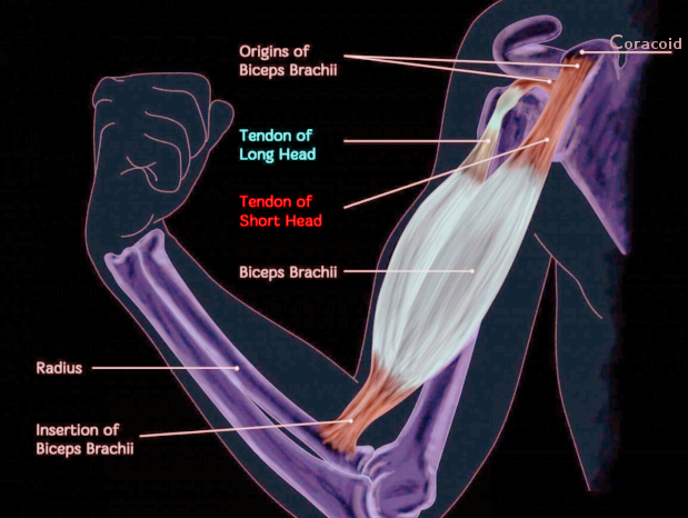
Biceps anatomy
Image courtesy O. Chaigasame
(Click Image to Enlarge)

Biceps tendon rupture
Image courtesy O Chaigasame