Physiology, Brain
- Article Author:
- Kenia Maldonado
- Article Editor:
- Khalid Alsayouri
- Updated:
- 5/24/2020 7:29:38 PM
- For CME on this topic:
- Physiology, Brain CME
- PubMed Link:
- Physiology, Brain
Introduction
The brain is an organ of nervous tissue that commands task-evoked responses, senses, movement, emotions, language, communication, thinking, and memory.
The parts of the human brain are:
- The cerebrum: It divides into the left and right cerebral hemispheres. The cerebral hemispheres have folds and convolutions on their surface. The ridges found between the convolutions are called gyri. The valleys between the gyri are called sulci (plural of sulcus). If the sulci are deep, they are called fissures. Both cerebral hemispheres have an outer layer of grey matter called the cerebral cortex.
- Cerebellum: It is comprised of the cerebellar cortex and deep cerebellar nuclei. The cerebellar cortex is made up of 3 layers, the molecular, Purkinje, and granular layers. The cerebellum is connected to the brainstem by structures known as the cerebellar peduncles. The cerebellum's primary function is to modulate motor coordination, posture, and balance.
- The brainstem: It contains the midbrain, pons, and medulla. It is located between the base of the cerebrum and the spinal cord.
Issues of Concern
Studies of brain function have focused on analyzing the variations of the electrical activity produced by the application of sensory stimuli. It is also essential to study the other features of the brain, including information processing and responding to environmental demands.[1]
The brain works precisely, making connections and is a deeply divided structure that has remained not entirely explained and even not examined.[2] Although researchers have made significant progress in experimental techniques, the human cognitive function that emerges from neuronal structure and dynamics is not entirely understood.[3]
Cellular
At the beginning of the forebrain formation, the neuroepithelial cells undergo divisions at the neural tube's inner surface to generate new progenitors. These dividing neuroepithelial cells transform and diversify, leading mostly to radial glial cells (RGCs).
RGCs also work as progenitors with the capacity of regenerating themselves, as well as the production of other types of progenitors, neurons, and glial cells.[4] They have long processes that connect with the neuroepithelium; they function as a guide for the migration of neuron cells so that neurons can find their resting place, mature and send out axons and dendrites to participate directly in synapsis and electrical signaling. Neurons get produced along with glial cells; the latter, in turn, bring support and create an enclosed environment in which neurons can perform their functions.
Glial cells (astrocytes, oligodendrocyte, microglial cell) roles that are well known include: keeping the ionic medium of neurons, controlling the rate of nerve signal propagation and synaptic action by regulating the uptake of neurotransmitters, providing a platform for some aspects of neural development, and also helping in recovery from neural damage.
Grey matter is the main component of the central nervous system (CNS); it consists of neuronal cell bodies, dendrites, myelinated and unmyelinated axons, glial cells, synapsis, and capillaries. The cerebral cortex is made up of layers of neurons that constitute the gray matter of the brain. The subcortical (beneath the cortex) area is mostly white matter, its major component is myelinated axons but just a few quantities of cell bodies compared to grey matter.
Although neurons can have different morphologies, they all contain four common regions: the cell body, the dendrites, the axon, and the axon terminals, and each one has its respective functions.
The cell body has a nucleus where proteins and membranes get synthesized. These proteins travel through the microtubules down to the axons and the terminals; it is called anterograde transport. Dendrites can also make some proteins, but the axon and terminal axons do not have ribosomes, so they cannot. Damaged membranes and organelles travel from the axon toward the cell body along axonal microtubules; this process is called retrograde transport, lysosomes are only present in the cell body, they contain and degrade the damaged material that reaches there. The axon is a very thin continuation of a neuron, whereby this cell sends nerve impulses to other types of cells.
Astrocytes occupy 25% of the total brain volume and are the most abundant glial cells.[5] These classify into two main groups: protoplasmic and fibrous. Protoplasmic astrocytes appear in the gray matter and have several branches that contact both synapses and blood vessels. Fibrous astrocytes are present in the white matter and have long fiber-like processes that contact the nodes of Ranvier and the blood vessels. Astrocytes use their connections to vessels to titrate blood flow in the response of synaptic activity. Astrocytic endfeet, which form tight junctions between endothelial cells and basal lamina together give rise to the formation of the blood-brain barrier (BBB).[6]
The primary function of oligodendrocytes is to make myelin; they are critical to maintaining the electrical impulse conduction and maximizing velocity; it is located in segments separated by nodes of Ranvier. Their function is equivalent to those of Schwan cells in the peripheral nervous system.
The macrophage populations of the CNS include the microglia, perivascular macrophages, meningeal macrophages, macrophages of the circumventricular organs (CVO) and the microglia of choroid plexus. Microglia are phagocytic cells that represent the immune and the support system of the CNS, and it is the most abundant and best studied of choroid plexus.[7]
Development
Human brain development starts with the neurulation process from the ectodermic layer of the embryo, and it takes, on average, 20 to 25 years to mature.[8] It occurs in a sequential and organized manner beginning with the neural tube formation at the third or fourth week of gestation and then followed by cell migration and proliferation that contribute to the increase in size and surface, creating a more complex structure. At birth, the general architecture of the brain is mostly complete, and by the age of 5 years, the total brain volume is about 95% of its adult size. Generally, the white matter increases with age, while gray matter decreases with age.
The most prominent white matter structure of the brain, the corpus callosum, increases approximately 1.8% per year between the ages of 3 and 18 years.[9] It conjugates the activity of the right and left hemispheres and its development impact on the progress of higher-order cognitive abilities.
Gray matter in the frontal lobe undergoes continued structural development reaching its maximal volume at 11 to 12 years of age before slowing down during adolescence and early adulthood. The gray matter in the temporal lobe follows a similar development pattern, reaching its maximum size at 16 to 17 years with a slight decline after that.[10]
Organ Systems Involved
The brain and the spinal cord comprise the CNS. The peripheral nervous system (PNS) subdivides into the somatic nervous system (SNS) and the autonomous nervous system (ANS). The SNS consists of peripheral nerve fibers that collect sensory information to the CNS, as well as motor fibers that send information from the CNS to skeletal muscle. The ANS function is to control the smooth muscle of the viscera and glands, and it consists of: the sympathetic nervous system (SNS), the parasympathetic nervous system (PNS), and the enteric nervous system (ENS).
The nerves from the brain connect with the different parts of the head and body, leading to different voluntary and involuntary functions. The ANS drives basic functions that control unconscious activities such as breathing, digestion, sweating, and trembling.
ENS provides the intrinsic innervation of the gastrointestinal system, and it is the most neurochemically diverse branch of the peripheral nervous system.[11] Neurotransmitters such as norepinephrine, epinephrine, dopamine, and serotonin have recently been a topic of interest due to their roles in gut physiology and CNS pathophysiology. They help in regulating gut blood flow, motility, and absorption.[12]
Function
The cerebrum takes control over the motor and sensory information, as well as conscious and unconscious behaviors, feelings, intelligence, and memory. The left hemisphere is responsible for controlling speech, and abstract thinking (the ability to think about things that are not actually present), while the right hemisphere is in charge of spatial thinking (thinking that finds meaning in the shape, size, orientation, location, the direction of objects, processes or phenomena).
The motor and sensory neurons descending from the brain cross to the opposite side in the brainstem. This crossing means that the right side of the brain controls the motor and sensory functions of the left side of the body, and the left side of the brain controls the motor and sensory functions of the right side of the body. Hence, a stroke affecting the left brain hemisphere, for example, will exhibit motor and sensory deficits in the right side of the body.
Sensory neurons bring sensory input from the body to the thalamus. The sensory information then gets relayed to the cerebrum through the thalamus. Hunger, thirst, and sleep are under the control of the hypothalamus.
The cerebrum is composed of four lobes:
- Frontal lobe: Is responsible for motor function, language, and cognitive processes, such as executive function, attention, memory, affect, mood, personality, self-awareness, and social and moral reasoning.[13] Broca area is located in the frontal lobe and is responsible for speaking and writing skills.
- Parietal love: Is responsible for interpreting vision, hearing, motor, sensory, and memory function.
- Temporal lobe: Wernicke area is located here, which is responsible for understanding spoken and written language. The temporal lobe is also an essential part of the social brain.[14] It processes sensory information for the retention of memories, language, and emotions. It also plays a major roll in hearing, spatial, and visual perception.
- Occipital lobe: This is the location of the visual cortex, and it interprets visual information.
The cerebellum controls the coordination of voluntary movement. It receives sensory information from the brain and spinal cord and fine-tunes the precision and accuracy of the motor activity. It also aids in cognitive functions such as attention, language, pleasure response, and regulation of fear.
The brainstem acts as a bridge that connects the cerebrum and cerebellum to the spinal cord. It has the principal centers to perform autonomic functions such as breathing, temperature, respiration, heart rate, wake-sleep cycles, coughing, sneezing, digestion, vomiting, and swallowing.
Mechanism
The brain represents 2% of the body weight. It consumes 15% of the cardiac output and 20% of total body oxygen. The resting brain consumes 20% of the body's energy supply. If the brain performs a task, the energy consumption increases an additional 5%. This fact shows that most of the brain's energy consumption gets used for intrinsic functions.
The brain uses glucose as its principal source of energy. During low glucose states, the brain utilizes ketone bodies as its primary source of energy. During exercise, the brain can use lactate as a source of energy.
In the developing brain, neurons follow molecular signals from regulatory cells like astrocytes to determine its location, the type of neurotransmitter it will secrete, and with which neurons it will communicate, leading to the formation of a circuit between neurons that will be in place during adulthood. In the adult brain, developed neurons fit in their corresponding place and develop axons and dendrites to connect with the neighboring neurons.[15]
Neurons communicate with each other via neurotransmitters released into the synaptic space, which is a 20 to 50-nanometer space between neurons. The neuron that releases the neurotransmitter into the synaptic space is called the pre-synaptic neuron, and the neuron that receives the neurotransmitter is called the postsynaptic neuron. An action potential in the presynaptic neuron leads to the influx of calcium and the subsequent release of neurotransmitters from their storage vesicle into the synaptic space. The neurotransmitter then travels to the postsynaptic neuron and binds to receptors to influence its activity. Neurotransmitters are rapidly removed from the synaptic space by enzymes.[16]
The oligodendrocytes in the CNS produce myelin. Myelin forms insulating sheaths around axons to allow the swift travel of electrical impulses through the axons. There are areas on the axons that have no covering with myelin called nodes of Ranvier. These nodes allow for sodium influx into the axon, which helps maintain the speed of the electrical impulse traveling through the axon. This transmission is called saltatory nerve conduction, which is the "jumping" of electrical impulses from one node to another and ensures that electrical signals do not lose their velocity and can propagate long distances without deterioration of the signal.[17]
Related Testing
Functional magnetic resonance imaging (fMRI) can track the effects of neural activity and the energy that the brain consumes by measuring components of the metabolic chain. Other techniques, such as single-photon emission computed tomography (SPECT) studies cerebral blood flow and neuroreceptors. Positron emission tomography (PET) assesses the glucose metabolism of the brain.[18]
Electroencephalography (EEG) records the brain's electrical activity and is very useful for detecting several types of brain disorders.
Advances in these techniques have enabled a broader vision and objective perceptions into mental disorders, leading to the improvement of diagnosis, treatment, and prognosis.
Pathophysiology
Injury to the brain stimulates the proliferation of astrocytes; this is an immunological response to neurodegenerative disorders called “reactive gliosis.”[19] Injury to neural tissue stimulates a series of molecular and morphological changes, being the most important the upregulation of the glial fibrillary acidic protein (GFAP). On the other hand, Epidermal growth factor receptor (EGFR) allows the transition from non-reactive to reactive astrocytes, and its inhibition improves axonal regeneration and rapid recovery, so when astrocytes are reactive, they proliferate and hypertrophy leading to glial scar formation.
The microglia represent the immune and support system of the CNS. They are neuroprotective in the young brain but can react abnormally to stimuli in the aged brain, and become neurotoxic and destructive, leading to neurodegeneration.[20] As the brain ages, microglia acquire a phenotype that is increasingly inflammatory and cytotoxic, generating a hazardous environment for neurons.[21] Hence, aging is the most critical risk factor in the development of several neurodegenerative diseases.
The brain is surrounded by cerebrospinal fluid and isolated from the bloodstream by the blood-brain-barrier (BBB). In cases like infectious meningitis and meningoencephalitis, acute inflammation causes a breakdown of the BBB, leading to the influx of blood-borne immune cells into the CNS. In other inflammatory brain disorders such as Alzheimer disease (AD), Parkinson disease (PD), Huntington disease (HD), or X-linked adrenoleukodystrophy, the primary insult is due to degenerative or metabolic processes, and there is no breakdown of the BBB.[22]
Oligodendrocyte loss can happen due to the production of reactive oxygen species or the activation of inflammatory cytokines, and this leads to the decreased production of myelin, leading to conditions such as multiple sclerosis (MS).[17]
Disturbances in the neurotransmitter systems are related to the production, release, reuptake, or receptor impairments of these substances and can cause neurologic or psychiatric disorders. Glutamate is the brain's most abundant excitatory neurotransmitter; meanwhile, GABA is the primary inhibitory transmitter in the brain. Glycine has a similar inhibitory action in the posterior parts of the brain. Acetylcholine helps in different processes like muscle stimulation at the neuromuscular junction (NMJ), digestion, arousal, salivation, and level of attention. Dopamine is involved in the reward and motivational component, motor control, and the regulation of the release of prolactin. Serotonin influences mood, feelings of happiness, and anxiety. Norepinephrine is involved in arousal, alertness, vigilance, and attention.
Cerebral oxygen delivery and consumption rate are ten times higher than global body values.[23] Blood glucose represents the primary source of energy for the brain, and the BBB is highly permeable for it. During low glucose states, the body has developed multiple ways to keep blood glucose within the normal range. As the level drops below 80 mg/dl, pancreatic beta-cells decrease insulin secretion to avoid further glucose decrease. If glucose drops further, pancreatic alpha-cells secrete glucagon and the adrenal medulla that releases epinephrine. Glucagon and epinephrine increase blood glucose levels. Cortisol and growth hormones also act to increase glucose, but they depend on the presence of glucagon and epinephrine to work.
Clinical Significance
Neurodegenerative Diseases
Neuronal degeneration worsens with age and can affect different areas of the brain leading to movement, memory, and cognition problems.
Parkinson's disease (PD) occurs due to the degeneration of the neurons that synthesize dopamine, leading to motor function deficits. Alzheimer's disease (AD) occurs due to abnormally folded protein deposition in the brain leading to neuronal degeneration. Huntington's disease occurs due to a genetic mutation that increases the production of the neurotransmitter glutamate. The excessive amounts of glutamate lead to the death of neurons present in the basal ganglia producing movement, cognitive, and psychiatric deficits. Vascular dementia occurs due to the death of neurons resulting from interruption of blood supply.
Although neurodegenerative diseases aren't classically speculated to be caused by disturbed metabolism, glucose metabolism has been suggested to be involved. Research has shown that there is a reduction in glucose metabolism in Alzheimer disease.[24]
Demyelinating Diseases
Demyelinating diseases result from damage to the myelin sheath that covers the nerve cells in the white matter of the brain, spinal cord, and optic nerves. Multiple sclerosis and leukodystrophies are a consequence of oligodendrocytes damage.
Stroke
A stroke occurs either when a blood vessel in the brain ruptures and bleeds or when a clot blocks a vessel. Hence, the blood supply to a specific part of the brain is lost, leading to neuronal death. This condition can result in one of several neurological problems depending on the affected region.
Brain Death
Neurologic determination of brain death is a complicated evaluation that may be misunderstood by nonspecialists and families.[25] Brain death is the complete and irreversible loss of brain activity, including the brainstem. It requires verification through well established clinical protocols and the support of specialized tests.
Hypoglycemia
Glucose is the primary source of energy for the maintenance of brain metabolism and function. The largest amount of it is used for information processing that is performed by networks of neurons.[26] The brain requires a continuous supply of glucose as it has limited glucose reserves. CNS symptoms and signs of hypoglycemia include focal neurological deficits, confusion, stupor, seizure, cognitive impairment, or death.
(Click Image to Enlarge)
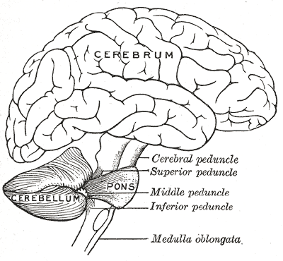
Brain, Encephalon, Connections of the several parts of the brain, Cerebrum, Cerebellum, Pons, Cerebral; Superior; Middle; Inferior Peduncle, Medulla oblongata
Contributed by Gray's Anatomy Plates
(Click Image to Enlarge)
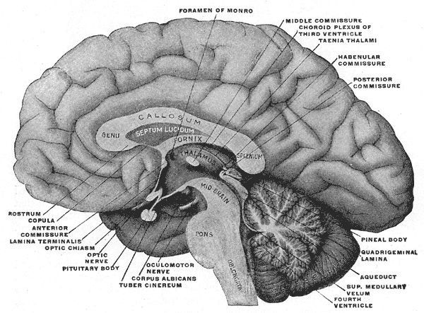
The Fore-brain or Prosencephalon, Mesal aspect of a brain sectioned in the median sagittal plane, Foramen of Monro, Middle commissure, Taenia thalami, Habenular commissure, Genu, Callosum, Fornix, Septum Lucidum, Plenum, Pons, Oblongata, Thalamus
Contributed by Gray's Anatomy Plates
(Click Image to Enlarge)
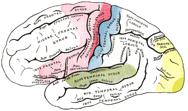
Areas of localization on lateral surface of hemisphere. Motor area in red, Area of general sensations in blue, Auditory area in green, Visual area in yellow, Brain, Neurology
Contributed by Gray's Anatomy Plates
(Click Image to Enlarge)

Pathways from the Brain to the Spinal Cord, The motor tract, Anterior nerve roots, Anterior and Lateral cerebrospinal Fasciculus, Decussation of pyramids, Geniculate fibers, Internal capsule, Motor area of cortex
Contributed by Gray's Anatomy Plates
(Click Image to Enlarge)
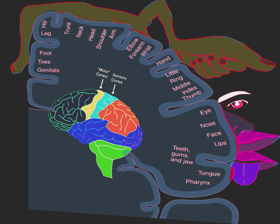
Homonculus: sensory & motor
Image courtesy S. Bhimji MD