Anatomy, Head and Neck, Carotid Sheath
- Article Author:
- Darren Garner
- Article Author:
- Michael Kortz
- Article Editor:
- Stephen Baker
- Updated:
- 7/1/2020 8:48:28 AM
- For CME on this topic:
- Anatomy, Head and Neck, Carotid Sheath CME
- PubMed Link:
- Anatomy, Head and Neck, Carotid Sheath
Introduction
The carotid sheath plays an important role in head and neck anatomy and contains several vital structures, including the carotid artery, jugular vein, vagus nerve, and sympathetic plexus. It arises in the base of the neck and terminates at the skull base. It is derived from mesoderm. While the carotid sheath itself is rarely the source of primary disease, understanding its anatomy is essential for clinicians to address problems that may affect its crucial components.
Structure and Function
The function of the carotid sheath is to separate and help protect the vital structures within it. It facilitates the passage of intrathoracic structures through the neck to terminate in the head and face. It is a fibrous connective tissue that encircles several key structures within the neck. These structures include:
- Common carotid artery
- Internal carotid artery
- Internal jugular vein
- Vagus nerve (CN X)
- Sympathetic plexus of nerves
The carotid sheath is located posterior to the sternocleidomastoid muscle and is a part of the deep cervical fascia of the neck. It consists of all three fascial layers of the neck, including the pretracheal fascia, the investing fascia, and the prevertebral fascia. The carotid sheath starts superior to the sternum and the first rib, and then extends to the base of the skull. Within the carotid sheath, the internal jugular vein runs lateral to the carotid artery with the vagus nerve posterior to both vessels in most individuals, though different anatomic configurations may exist and should be clinically accounted for.
Embryology
The carotid sheath, like other fascial tissues, is derived from mesoderm. The adventitia of the cervical great arteries becomes apparent by 15 weeks gestation and is one of the earliest components of the fetal deep cervical fascia. The carotid sheath does not appear until approximately 20 weeks gestation. At this time it is fused with the pretracheal fascia, but not yet integrated into the prevertebral lamina of the deep cervical fascia. [1][2][3][4]
Blood Supply and Lymphatics
There is no significant direct blood supply to the carotid sheath itself. The right common carotid artery originates at the bifurcation of the brachiocephalic artery, while the left common carotid artery arises from the aortic arch. Both common carotid arteries run within the carotid sheath until they bifurcate into the internal and external carotid arteries at the level of the upper border of the thyroid cartilage. After the bifurcation, the external carotid artery exits the sheath and supplies blood via eight main branches to various structures of the face and neck. The internal carotid artery continues within the carotid sheath and enters into the temporal bone through the carotid canal and gives rise to the ophthalmic artery as well as the anterior and middle cerebral arteries.
The internal jugular vein is a continuation of the sigmoid sinus and begins its descent towards the heart at the jugular foramen, running with the vagus nerve. It travels within the carotid sheath and drains into the subclavian vein, ultimately forming the brachiocephalic vein. While moving posteriorly, the internal jugular receives venous blood from the facial, lingual, and superior and middle thyroid veins.
Nerves
The carotid body and carotid sinus are located at the carotid bifurcation and function as important receptors within the body. The carotid body is a chemoreceptor that is sensitive to chemical changes including oxygen, carbon dioxide, and hydrogen ion concentration within the blood and helps control respiration. The carotid sinus is located just above the bifurcation at the origin of the internal carotid artery and functions as a baroreceptor helping detect and correct changes in blood pressure.
The vagus nerve has the longest course of any cranial nerve and begins its journey by passing through the jugular foramen with the internal jugular vein and makes it descent within the carotid sheath. Within the neck, it gives off the pharyngeal branch and the superior laryngeal nerve. The pharyngeal branch supplies motor fibers to the muscles of the pharynx with the exception of the stylopharyngeus. A lesion to the pharyngeal branch causes deviation of the uvula to the opposite side of the injury. The superior laryngeal nerve divides into the internal and external laryngeal nerves. These nerves supply sensory fibers to the larynx above the vocal cord, lower pharynx, and epiglottis as well as supplying taste fibers to the root of the tongue near the epiglottis.
The glossopharyngeal nerve (CN IX), accessory nerve (CN XI), and hypoglossal nerve (CN XII) pierce the superior section of the carotid sheath briefly and then exit. In addition, the ansa cervicalis is embedded in the anterior portion of the carotid sheath. It is important to visualize these structures when performing procedures that require incision or manipulation of the carotid sheath.
Muscles
As previously stated, the carotid sheath is derived from all three layers of the deep cervical fascia. While there are no muscles contained within the carotid sheath itself, the sternocleidomastoid muscle does provide an anatomic landmark as its anterolateral boundary.
Physiologic Variants
There are no significant physiologic or anatomic variants of the carotid sheath, however, it can be disrupted or lay on an unusual fascial plane if the common carotid arteries have a unique course through the neck. Carotid variations are fairly common (i.e. the right common carotid branching off the aorta directly). Thus, access to the carotid sheath's contents should be performed with care and can be done under the aid of ultrasound or other imaging if an anatomic variation is suspected.
Surgical Considerations
Carotid Endarterectomy
A carotid endarterectomy is performed to excise atherosclerotic thickening of the intima within the internal carotid artery in an effort to reduce strokes in patients with significant carotid artery stenosis (typically >70-80% stenosis +/- symptoms). To begin, the opening incision is made and the sternocleidomastoid muscle is retracted. At this point it is important to visualize the carotid sheath for this is where the structure vital to the procedure is located. The carotid artery lies on the medial side of the internal jugular vein, and the vagus nerve is situated posteriorly. Superiorly, the carotid sheath may also contain the hypoglossal nerve, the glossopharyngeal nerve, and the accessory nerve. These structures pass in a horizontal fashion and cross the internal carotid artery. It is important to identify these structures before incising any structure. The surgeon opens the carotid sheath to gain exposure to the common carotid bifurcation, as this is the most common site for atherosclerosis due to non-laminar blood flow. Once access is gained to the carotid sheath and the carotid bifurcation is located, the surgeon then removes any atherosclerotic plaque found and repairs the vessel. Possible complications include air embolism and laceration of the internal jugular vein or carotid artery.[5][6][7]
Penetrating Neck Trauma
An ongoing debate among trauma surgeons is over a no-zone approach, which leans heavily on multidetector CT angiography versus a zoned approach to surgical exploration.[8][9]
Zones of the Neck
The neck is divided into three zones. These become important when assessing and managing trauma in those with neck injuries.
Zone I - cricoid cartilage to the sternal notch: trachea, lung, esophagus, thoracic duct, vertebral arteries, origin of the common carotid artery, and subclavian vessels, spinal cord, thoracic duct, thyroid gland
Zone II - cricoid cartilage to angle of mandible: carotid sheath and its components (carotid artery, internal jugular vein, vagus nerve), trachea, esophagus, spinal cord, larynx, pharynx
Zone III - angle of mandible to base of skull: distal portion of internal carotid artery, vertebral arteries, jugular veins, pharynx, spinal cord, sympathetic chain, CN IX,X,XI,XII
The decision to explore these areas is based on hard and soft signs. Regardless of the zone, if the patient becomes unstable, surgical exploration is the best course of management.
Soft Signs
- Hypotension in field
- History of arterial bleeding
- Tracheal deviation
- Nonexpanding large hematoma
- Apical capping on chest radiograph
- Stridor
- Hoarseness
- Vocal cord paralysis
- Subcutaneous emphysema
- Seventh cranial nerve injury
- Unexplained bradycardia (without CNS injury)
Hard Signs
- Hypotension in Emergency Department
- Active arterial bleeding
- Diminished carotid pulse
- Expanding hematoma
- Thrill/bruit
- Neurologic deficit
- Hemothorax greater than 1,000 mL
- Air bubbling of wound
- Hemoptysis
- Hematemesis
If the patient presents with hard signs then emergent management is needed (i.e. airway and circulation management, immediate decompression, and repair of the injured vessel), whereas if soft signs are present then serial examination and non-emergent imaging may be appropriate.
Clinical Significance
Internal Jugular Central Venous Line
An internal jugular central line is performed to gain access to push medication or fluids into the systemic vasculature. The procedure begins by placing the patient in slight Trendelenburg and locating the internal jugular vein within the apex of the triangular interval between the clavicular and sternal heads of the sternocleidomastoid muscle. It may help to have the patient turn the head to the contralateral side to better visualize these anatomical landmarks. If the patient is conscious, begin by applying a local anesthetic. Next, insert the needle at a 45-degree angle posteriorly while applying negative pressure. The needle will pass through the carotid sheath and the internal jugular vein will produce blood within the syringe. An ultrasound can be used to help assist and ensure proper placement of the guide needle into the vein. While keeping the guide needle in place, begin threading the guide-wire. Once in place, remove the needle and introduce the dilator and then the catheter. The catheter should now be within the superior vena cava.
It is important to understand the relationships of the structures within the carotid sheath to properly perform an internal central venous line.
Lemierre Syndrome
Lemierre Syndrome, otherwise known as suppurative jugular thrombophlebitis, is a serious sequela of odontogenic or oropharyngeal infection. Infection typically spreads from the lateral pharyngeal space to the carotid sheath. Here, it can seed the internal jugular vein and precipitate septicemia and septic emboli, as well as erode the carotid artery. It is typically due to Fusobacterium or Bacteroides species. While it is uncommon in the antibiotic era, it can still present in patients with poor dental care and access to the healthcare system.
Other Issues
The carotid sheath should not be confused with the carotid space or carotid triangle. The carotid space is the anatomic area that the sheath encloses, while the carotid triangle is a specific portion of the anterior aspect of the neck in which the carotid sheath passes through.
(Click Image to Enlarge)
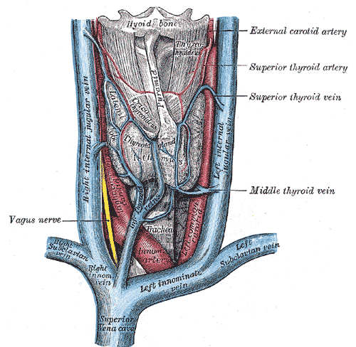
Veins and Arteries of the neck, Superior Vena Cava, Left and Right Innominate vein, Left and Right Subclavian vein, Left and Right Internal jugular vein, Innominate Arteries, Hyoid Bone, Thyroid Gland, Trachea
Contributed by Gray's Anatomy Plates