Facial Nerve Trauma
- Article Author:
- Rakesh Mistry
- Article Editor:
- Ahmed Al-Sayed
- Updated:
- 8/10/2020 9:28:53 PM
- For CME on this topic:
- Facial Nerve Trauma CME
- PubMed Link:
- Facial Nerve Trauma
Introduction
Facial nerve palsy is a common presentation to primary care providers, the emergency department, and otolaryngologists. Trauma accounts for 10% to 23% of all facial nerve palsies. It has implications on a patient’s quality of life due to the role the facial nerve has in multiple important processes. Alongside the emotional impact of facial expression, facial nerve palsy can have ophthalmological, otological, rhinological, taste, and swallowing consequences. Ophthalmological consequences from impaired lacrimation, ectropion, and epiphora can lead to exposure keratopathy, which can lead to blindness. Otological consequences include a reduction in hearing. Impaired muscular support to the nasal valve can lead to nasal obstruction. Impact on the perioral musculature can result in poor swallowing and insufficient oral competence, while the damage to the chorda tympani branch of the facial nerve can lead to impaired or altered taste.[1]
Etiology
Trauma to the facial nerve has been described in the following categories [1]:
- Basal skull fracture (temporal bone)
- Penetrating trauma to the extratemporal aspect of the facial nerve
- Birth (forceps delivery)
- Iatrogenic (for example post parotidectomy or mastoidectomy)
- Barotrauma (altitude paralysis or scuba diving)
- Lightning
This activity focuses on the most common cause of trauma, leading to facial nerve palsy, which being basal skull fracture.[2]
Epidemiology
The facial nerve suffers injury in 7% to 10% of all temporal bone fractures. The pyramidal shape of the temporal bone means a significant force is necessary to break the temporal bone. Therefore, 31% of temporal bone fractures occur secondary to motor vehicle collisions. Following this, the next most common mechanisms are assault or falls.[1][3] Consequently, approximately 90% of temporal bone fractures are associated with intracranial injuries, and 10% are associated with cervical spine injuries.[4]
Pathophysiology
The nature of the temporal bone fracture is useful in determining the likelihood of involvement of the facial nerve as well as potential complications.
The Ulrich classification of temporal bone fractures was suggested in 1926 and is dependent on the orientation of the fracture to the petrous ridge of the temporal bone [3]:
Longitudinal (approximately 75%)
These are along the temporal bone longitudinal axis and associated with temporoparietal trauma (lateral blow).
These fractures rarely involve the otic capsule as the fracture line follows the path of least resistance towards the petrous apex of the petrous temporal bone.
However, these fractures tend to result in ossicular injury (ossicular chain disruption), haemotympanum, tympanic membrane perforation, and otorrhagia. Alongside this is an associated conductive hearing loss.
Facial nerve palsy is associated with 20% of longitudinal temporal bone fractures.[3]
Transverse (approximately 20%)
These are perpendicular to the long axis of the petrous bone and through the labyrinthine capsule and have correlations with frontoparietal trauma (front-back trauma).
They usually originate around the jugular foramen or foramen magnum and extend into the middle cranial fossa. Consequently, these fractures commonly result in a sensorineural hearing loss, which may be a result of transection of the cochlear nerve, damage to the labyrinthine structures, or stapes footplate injury.
They are associated with perilymphatic fistula (with nystagmus away from the fractured side) and facial palsy in 50% of cases.[3]
Mixed
This condition is a combination of transverse and longitudinal.
These commonly involve the otic capsule, which can lead to sensorineural hearing loss. If there is an ossicular injury, there will be an element of conductive hearing loss as well.
However, recent studies have found that the majority of fractures do not easily fit into these categories, and the scheme does not provide useful prognostic information concerning neurotologic deficits. Hence a new classification was suggested. The reason for mentioning the above classification is that it is still commonly used in practice when describing a temporal bone fracture.
Brodie et al. suggested a classification distinguishing fractures as otic sparing versus otic capsule disrupting. The classification has information regarding functional sequelae and, consequently, prognosis.[2][3]
Otic Capsule Disrupting fractures (2.5 to 5.8%)
Usually caused by occipital blows, with the fracture line running from foramen magnum across the petrous pyramid to the otic capsule.
Commonly they pass through the jugular foramen, internal auditory canal, and foramen lacerum.
Consequently, they are associated with the following complications:
- Facial nerve palsy (2 to 5 times as likely)
- Cerebrospinal fluid (CSF) leak (4 to 8 times as likely) and fistula
- Sensorineural hearing loss (7 to 25 times as likely)
- Epidural hematoma and subarachnoid hemorrhage
Otic Capsule Sparing fractures
Usually caused by damage to the temporoparietal region and involve the squamous temporal bone and posterosuperior wall of the external auditory canal.[5]
History and Physical
It is essential to determine the onset and progression of facial palsy in traumatic circumstances as research demonstrates the onset of facial paralysis has an impact on prognosis and management. Immediate complete facial paralysis is the most important prognostic factor. These patients typically have much more severe facial nerve injuries and are more likely to benefit from facial nerve exploration and repair. These patients tend to have a poor prognosis.
Patients who present with normal or incomplete facial paralysis will rarely require facial nerve decompression and exploration. A partial facial nerve injury can progress to a complete paralysis over a few days (due to swelling causing compression of the nerve in the facial canal). Patients who present with a paresis rather than paralysis, which later progress to complete paralysis, generally have a good prognosis for spontaneous recovery.
Hence, early clinical evaluation to establish baseline facial nerve function is critical.
Immediate assessment of the facial nerve could prove particularly challenging in the setting of polytrauma, especially when administering paralytic agents for intubation purposes. Sometimes the exam is omitted to focus on other life-threatening issues. It is crucial to distinguish "delayed onset" facial palsy from "delayed diagnosis" of facial palsy. Delayed onset facial palsy is defined as a facial nerve function that subsequently deteriorates from initial presentation, whereas delayed diagnosis occurs when facial nerve function is not assessable. In this situation, these patients should be categorized as unestablished onset facial palsy and treated as immediate onset facial palsy.
Evaluation
Clinical Assessment
Given the potential for a high impact injury, a thorough history and examination are vital to ensure the examiner does not miss other intracranial or spinal injuries. A systematic ABCDE approach, according to Advanced Trauma and Life Support (ATLS) guidelines, should be followed.
Clinical examination should include a full neurological assessment and otoscopy. The patient should undergo evaluation for evidence of basal skull fracture; this should include assessment for raccoon eyes and battle signs as well as hemotympanum or bloody otorrhea and any evidence of CSF leak.
The functional status of the facial nerve is recorded as soon as possible and provides valuable prognostic data. This function assessment should use the House-Brackmann scale, which is a useful tool, especially in the hands of experienced clinicians [6]:
- I - Normal symmetrical function throughout
- II - Slight weakness on close inspection + slight asymmetry of smile
- III - Obvious non-disfiguring weakness, complete eye closure with maximal effort
- IV - Obvious disfiguring weakness, cannot lift the brow, incomplete eye closure, severe synkinesis
- V - Barely perceptible motion, incomplete eye closure, a slight movement of corner of the mouth, absent synkinesis
- VI - No movement, atonic
Hearing requires assessment. In the acute setting, this is most likely with whisper test and tuning forks) to assess the nature of any hearing loss. If possible, the clinician should obtain audiometry.
Any material suspicious for CSF should undergo evaluation for beta-2 transferrin.
Radiological Assessment
The majority of trauma patients will undergo a trauma CT series, which can provide sufficient details of any temporal bone fracture. If there is an acute onset of facial nerve paralysis or CSF leak, then a high-resolution CT (HRCT) of the temporal bones may be requested.[3]
Nerve Function Testing
These tests are best at least 2 to14 days after injury for Wallerian degeneration of axons to take place.[1] They are only necessary in cases of paralysis. It is not used in cases of paresis as the nerve is intact.
The two types of tests are:
- Electroneurography or evoked electromyography (ENOG or evoked EMG) - measures evoked muscle action potential using skin electrodes. Nerve injury is a percentage of function relative to the normal side.
- Electromyography (EMG) - measures voluntary muscle response with electrodes in the target muscle detecting action potentials during muscle contraction with a functioning nerve.
When ENOG is undetectable, EMG is an option.
The above tests can help classify the nerve injury according to the Sunderland classification and subsequently, direct management [7]:
- Conduction block (neuropraxia)
- Axonal injury (axonotmesis)
- Type 2 + endometrium injury (neurotmesis)
- Type 3 + perineurium injury (neurotmesis)
- Type 4 + epineurium injury (neurotmesis)
The above tests are only useful in distinguishing injuries that do not cause Wallerian degeneration (type 1) from those that do (type 2-5).
If there is a greater than 90% degeneration within six days of the injury or greater than 95% degeneration within 14 days, there is likely to be a Sunderland type 5 injury with poor prognosis.[3]
Treatment / Management
There has been significant debate surrounding the selection of patients for surgery in facial nerve trauma. The trouble with the literature comparing facial nerve decompression (surgery) versus conservative management is the quality of studies and small amounts of studies. Consequently, there is no evidence to prove or disprove facial nerve decompression. One paper demonstrated that conservative, nonoperative management is associated with facial nerve recovery in 63% of immediate-onset complete facial nerve paralysis compared to 51% that underwent facial nerve decompression.[3] This recovery can take up to 1 year even after surgery, with some degree of residual synkinesis and muscle weakness in some cases.
The decision for surgical intervention is likely to be based on whether the injury to the nerve is likely to be neuropraxia (i.e., recoverable) or severed/crushed (i.e., unrecoverable).
The key to the management is identifying acute onset, a delayed onset, and those that had a late diagnosis.
There can be difficulty in categorizing patients. For example, as a patient following a road traffic collision may be intubated and sedated due to other injuries. These patients should be treated as immediate-onset facial paralysis to avoid inappropriate conservative management.
Therefore the below details are a general rule for treatment based on literature. However, local experts will guide the decision of surgical intervention.
Medical
High dose corticosteroids are appropriate in cases of delayed onset complete paralysis or incomplete paresis. These patients generally demonstrate good prognosis. The rationale is based on reducing neural edema, which is causing the progression of injury of the non-transected nerve.
Prophylactic antibiotics for CSF leak following a recent meta-analysis dedicated to CSF leaks relating to the lateral skull base.[3]
Eyecare is incredibly essential in those with palsies resulting in corneal exposure. Artificial tears, adequate lubricant, and taping the eyes closed at night ensure the prevention of corneal ulceration. Ophthalmology referral is essential.
Surgical
If there is immediate complete paralysis, then surgical exploration for facial nerve decompression is required as soon as the patient’s condition allows; this is usually within 2 to 3 weeks, although this is debatable.[2][3]
If there is a delayed diagnosis of paralysis with neural degeneration of 90% or more on ENOG, then surgical decompression is also indicated. Some literature suggests the use of a combination of electrodiagnostic and CT, especially if the timing of the palsy is not known.
If facial nerve decompression is the desired option, then the approach and timing must also be considered. Most commonly, the facial nerve suffers injury at the perigeniculate region and the mastoid segment. Hence these must be exposed.[3]
A transmastoid/supralabyrinthine approach avoids the sacrifice of sensorineural hearing and intracranial exposure. However, it requires dislocation of the incus and reconstruction of the ossicles. Therefore, this approach is utilized in otic capsule sparing fractures with ossicular discontinuity. These fractures tend to be distal to the geniculate ganglion.[2][3]
For example, if the clinician suspects the patient has a fracture involving the mastoid or squamous portion (often those with Battle sign), then the fracture line is followed medially until the point of facial nerve injury. If the line is not present, the facial nerve should be exposed in the facial canal and traced along until detecting the damage. The incus requires removal to complete this.[2]
If the injury is proximal to the geniculate ganglion with no sensorineural hearing loss, the anatomy is not conducive to this approach, or the patient has contralateral hearing loss, then a middle cranial fossa approach is utilized. This approach requires the creation of a temporal bone flap parallel to the middle cranial fossa. Careful dissection is requisite to prevent bleeding from the middle meningeal artery, which can be embedded in the inner table. Care is necessary to avoid the superior semicircular canal and basal turn of the cochlea. Temporalis fascia serves to close the dural defect at the internal acoustic meatus to prevent temporal lobe herniation into the middle ear.[2][3]
A translabyrinthine approach is useful in patients in otic capsule disruption fractures. Preexisting profound sensorineural hearing loss is unlikely to improve. This approach has the advantage of excellent exposure to almost the entire course of the intratemporal facial nerve. Direct re-anastomosis and cable grafting can then follow. The closure is with abdominal fat grafts to maintain water-tight closure to minimize CSF leakage.[2][3]
The timing of any surgical intervention is also a topic of debate. Some literature advocates early intervention to prevent further degeneration, which is within seven days after acute-onset, complete paralysis; however, others suggest if the nerve does not undergo repair within three days, then to delay decompression for 20 days after the injury as axoplasmic flow and regeneration are the greatest three weeks after injury.[3]
The options to manage the nerve injury depend on the operative findings:
Nerve damage secondary to impingement (the nerve is intact)
Decompression of epineural sheath in a proximal to distal fashion.
This procedure is more common and is often due to things such as stretch trauma, impingement by bony spicules, or intraneural hematoma. This surgery often gets delayed due to the treatment of life-threatening injuries suffered in the original trauma, delayed referral, or poor neurological status. This delay does not worsen their prognosis, with surgery shown to still be of benefit three months following injury.
Nerve transection (Sunderland 5th degree of nerve injury)
Either primary neurorrhaphy using fine suture or fibrin glue or an interposition nerve graft when a primary anastomosis is not possible
Interpositional nerve graft is common with the greater auricular nerve. Patients who have surgery can expect a House–Brackmann grade III or better after a 2-year follow-up, with just under half of these patients demonstrating near-normal function.[1]
Surgical Management of Facial Paralysis
As with all surgical interventions, the treatment plan must tailor to the individual patient. The plan must take into account their age, comorbidities, and etiology, as well as the severity of the paralysis. A large number of facial reanimation surgical procedures are lengthy and, therefore, not suitable to the elderly or those with significant comorbidities due to the high risk of perioperative morbidity and mortality. The duration of paralysis is essential as research demonstrates reinnervation procedures are less effective after two years as there is the complete degradation of motor endplates.[1]
There remains a stepwise approach for surgical management of facial nerve palsy, which includes primary neurorrhaphy, interposition nerve graft, cross-facial nerve graft (CFNG), nerve transposition, regional muscle transfer, and free-tissue transfer.
If possible, the clinician should attempt primary nerve repair. The only exception to this is cases where the facial paralysis has been long-standing with degeneration of motor endplates.
The decision between dynamic reconstruction and static repositioning depends on the timeframe of paralysis. In cases of acute trauma (for example, nerve sectioning or penetrating wounds), then primary repair has demonstrated the best outcomes. In particular, a tension-free coaptation (instead of tension coaptation) allows improved perfusion and neural regeneration.
When primary neurorrhaphy is not possible, an interposition nerve graft is the option of choice. The most common source is the great auricular or sural nerves. The coaptation of the nerve should be in the portion of the facial nerve distal to the stylomastoid foramen as the nerve fibers proximal to the stylomastoid foramen are less favorable and can lead to greater synkinesis.[1]
When primary neurorrhaphy or interposition nerve graft is not possible, and if the patient is less than two years from injury, the surgeon can attempt CFNG, which involves utilizing the peripheral branches of the contralateral intact facial nerve to innervate the paralyzed hemiface. It is suitable for those within two years from injury because the motor end plates remain intact, and the facial musculature is less likely to have atrophied by this point.
Due to the time taken for the axons to travel to their target, this technique does risk ongoing muscle atrophy. Therefore, in patients who are more than six months since their injury, a ‘babysitter’ procedure may be utilized. This process involves a partial hypoglossal nerve transfer alongside the CFNG to provide more rapid neural input. This procedure has resulted in a near symmetrical smile in 65% and a symmetrical smile in 10% in one study.[8]
Nerve transposition utilizes the ipsilateral hypoglossal (most common), masseteric, spinal accessory, ansa cervicalis, or recurrent laryngeal nerve fibers. It is an option when CFNG is not an option. Hypoglossal nerve transfer can use either the superior or inferior halves of the nerve and gives reanimation to a House-Brackmann grade 2 to 6.[9]
Regional muscle transfer most commonly involves the temporalis muscle, which can be via regional muscle transposition involving rotation of temporalis over the zygomatic arch and anchoring to the oral commissure. An alternative may be temporalis tendon transfer via a transbuccal or nasolabial fold incision. The tendon transfer has the advantage of avoiding excessive temple depression and tissue density in the region of the zygomatic arch that is associated with regional muscle transposition.[10]
A free-tissue transfer can take place utilizing a latissimus dorsi or gracilis free flap. The latissimus dorsi flap involves immediate coaptation of the thoracodorsal nerve to the contralateral nerve, whereas the gracilis free flap involves innervation to the ipsilateral masseter nerve. Although the gracilis flap has demonstrated greater dynamic displacement of the oral commissure and increased conduction velocity compared to CFNH techniques, one study found that latissimus flaps provided voluntary and spontaneous smiling in 92.5% of cases. In contrast, gracilis flaps demonstrated spontaneous smile in only 10%.[1]
When considering treatment, the face can undergo segmentation into three facial zones – upper, middle, and lower. Treatment can involve the affected side, the contralateral, or both.
Upper Face
The focus of the upper face is (a) protection of the eye and (b) brow symmetry.
Exposure keratopathy secondary to ectropion in facial paralysis is caused by corneal desiccation through lagophthalmos and lacrimal dysfunction.[11] It can be avoided through lubricating eye drops/ointments and taping the eye shut at night. An alternative may be botulinum toxin injected into Muller's muscle and levator palpebrae superioris (to counter lagophthalmos) or hyaluronic acid into the upper lid (to promote eye closure). Lateral ectropion can also be addressed surgically with a lateral tarsal strip or lateral transorbital canthopexy. Medial ectropion is addressable via medial canthopexy. The approach could be transcutaneous, transcaruncular, or precaruncular.
Static weight implantation into the upper eyelid using either gold or platinum is an option to help eye closure. One study demonstrated that gold implantation alongside lateral tarsorrhaphy provided complete eye closure in 83% after one procedure.[1] Fourteen percent of patients, however, required a revision procedure to optimize lid weight. Platinum has demonstrated to provide a more significant benefit due to higher density and better biocompatibility. One study of 1000 patients showed reduced capsule formation and extrusion compared to gold.[12]
When considering dynamic reanimation of the eye, this is possible by nerve transfers or muscle transfers. There is some evidence suggesting better results for nerve transfers.[13] Nerve transfer can be with cross-facial nerve grafting or by hypoglossal nerve transfer. Muscle transfer procedures would involve frontal, temporalis, or free muscle flaps.
Exacerbation of dermatochalasis of the brow due to brow ptosis can obscure the upper visual field; this is treatable with brow elevation using open or endoscopic techniques. The endoscopic approach has demonstrated faster recovery as well as reduced sensory disturbance and alopecia.[1]
Midface
The treatment aim of the mid-face is to reduce/relieve nasal obstruction.
Symptomatic stenosis of the nasal valve is treatable with a minimally invasive suture suspension or polytetrafluoroethylene sling to suspend the nasolabial fold.[14][15] Other options that have improved nasal airway flow are rhytidectomy or functional septorhinoplasty.[1]
Paralysis of the midface can have an impact on both nasal valve stenosis but also exacerbation of ectropion. The clinician can address this through techniques such as an extended minimal access cranial suspension lift or mobilization of the suborbicularis oculi fat and periosteum.
Lower Face
The treatment aims for the lower face focus on preventing ptyalism, difficulty eating and drinking, poor articulation, and loss of facial expression.
For patients who are poor surgical candidates or have had long term facial paralysis, static slings constructed from fascia lata or polytetrafluoroethylene are a viable option. These create a nasolabial fold through suspension from the zygomatic arch or deep temporal fascia. A face-lifting procedure that achieves a similar result is a minimal access cranial suspension. Lower lip reanimation is achievable via palmaris longs tendon transfer.[1]
Non-Operative Approach
For patients who are not suitable surgical candidates, neuromuscular facial retraining can be implemented. This process utilizes biofeedback and focuses on the patient practicing using a mirror or EMG.
Adjunctive therapy may be the injection of botulinum toxin. The toxin gets injected on the normal side of the face, which weakens it while also strengthening the paralyzed half resulting in improved symmetry. One study reported beneficial effects were noted at six months after the pharmacological effects of the toxin had completed.[1]
Differential Diagnosis
Other causes of facial nerve palsy are:
1. Bells palsy
2. Ramsay Hunt syndrome
3. Cerebrovascular accident
4. Acoustic neuroma
5. Parotid malignancy
6. Malignant otitis externa
7. Otitis media
These can often be ruled out through careful history and examination findings.
Prognosis
As detailed above, there is debate surrounding the management of these patients and, subsequently, prognosis. One paper demonstrated that conservative, nonoperative management is associated with facial nerve recovery in 63% of immediate-onset complete facial nerve paralysis compared to 51% that underwent facial nerve decompression.[3] This recovery can take up to 1 year even after surgery, with some degree of residual synkinesis and muscle weakness in some cases.
Complications
Complications of facial nerve palsy are detailed in the introduction and include emotional and psychological harm alongside ophthalmological, otological, rhinological, and impact on taste.
Complications of temporal bone fractures include CSF leak, meningitis, CSF fistulae, cholesteatoma formation, external auditory canal stenosis, and intratemporal carotid injury.[3]
Consultations
1- Neurosurgical consultation for intracranial complications
2- Ophthalmological consultation for ophthalmic complications
3- Facial plastic surgeons for facial reanimation or functional static procedures
4- Physiotherapists for rehabilitation
5- Electrophysiologists for nerve conduction studies and EMG
Deterrence and Patient Education
Unfortunately, trauma sometimes cannot be avoided. Therefore, patient education revolves around ensuring the patient has been informed around the signs of facial nerve palsy if it were to present delayed following the trauma.
Pearls and Other Issues
Anatomy: Facial Nerve
The course of the facial nerve divides into three major segments - origin, intratemporal, and extratemporal
The origin of the facial nerve can subdivide into the intracranial and metal segments. Within the intracranial and meatal segments, there are no branches given off.
The intracranial segment
The motor nucleus of the facial nerve originates within the lower pons and emerges via the cerebellopontine angle. It is then joined by the nervus intermedius, which consists of the sensory and autonomic fibers of the facial nerve, which originate from the tractus solitarius and superior salivatory nucleus, respectively.
The meatal segment
The facial nerve then inserts into the internal acoustic meatus (IAM) to begin its meatal segment. The IAM is in the petrous part of the temporal bone. Within the IAM, the facial nerve runs in the anterosuperior compartment.
Intratemporal
The intratemporal segment subdivides into the labyrinthine, tympanic, and mastoid segments.
Labyrinthine segment
The labyrinthine segment runs between the IAM+ the geniculate ganglion/first Genu and is the narrowest portion of the facial nerve. Hence, it is most vulnerable to compromise at this point. At the level of the geniculate ganglion, the facial nerve undertakes the first of two sharp bends (first genu).
Here, the greater petrosal nerve branches off from the main trunk. The greater petrosal nerve provides preganglionic parasympathetic fibers to the pterygopalatine ganglion (also known as the vidian nerve). The pterygopalatine ganglion provides postganglionic parasympathetic fibers to the lacrimal, nasal and palatine glands. Proximal lesions are associated with impaired lacrimation, hyperacusis, and loss of taste on the anterior two-thirds of the tongue.
Tympanic segment - exists between the geniculate ganglion and the second genu
The facial nerve moves through the fallopian canal before its second sharp bend (genu). Important relations of the facial nerve at this point include:
Anteriorly: Processus cochleariformis (where tensor tympani tendon gets directed to the malleus). Posteriorly: oval window (inferiorly) and the lateral semi-circular canal (superiorly).
Mastoid segment - runs from the second genu to the stylomastoid foramen.
After its second genu, the nerve runs on the posterior aspect of the tympanic cavity.
Both the nerve to stapedius and chorda tympani leave at this segment.
The chorda tympani runs anteriorly across the tympanic cavity to provide preganglionic parasympathetic fibers to the submandibular ganglion, which then provides parasympathetic innervation to the submandibular and sublingual glands. The nerve to stapedius innervates the stapedius muscle.
Extratemporal
The nerve moves inferiorly and laterally around the styloid process. Before entering the parotid gland, it gives off branches to the three following muscles:
- Occipitalis
- Stylohyoid
- Posterior belly of digastric
Within the parotid gland, the nerve divides the gland into superficial and deep parts. It lies the most superficial structure traversing the parotid gland.
Within the substance of the parotid gland, it divides into two trunks (cervicofacial and temporofacial) and then five main branches which each are responsible for innervation of the muscles of facial expression.
The five branches with their target muscle and action are:
Branch of facial nerve/Primary target muscle/Clinical assessment
Temporal/frontalis/raise eyebrows
Zygomatic/orbicularis oculi/close eyes
Buccal/puff cheeks out
Mandibular/depressor anguli oris/show bottom teeth
Cervical/platysma/clench neck
To summarize, the facial nerve innervates the following muscles:
- Stapedius
- Stylohyoid
- Posterior belly of digastric
- Occipitalis
- Muscles of facial expression
Anatomy: Otic Capsule
The otic capsule (bony labyrinth) is part of the dense bone of the petrous temporal bone that surrounds the membranous labyrinth of the inner ear. It consists of the cochlea, vestibule, and semicircular canals (all of which contain their membranous counterparts). Between the bony and membranous labyrinth sits the perilymph.[16]
Timing of Paralysis
Overall there is approximately a 6% to 7% risk of facial paralysis for patients with temporal bone fractures. The timing and nature of the paralysis are important as it guides management. One study found it to be immediate in 27% of cases and delayed in 73% (deemed at 1 to 16 days). Delayed onset is a documented facial function in the emergency room that subsequently deteriorates.
In one case series, there was immediate complete paralysis in 51% of cases, delayed complete paralysis in 15%, and paresis in 23% of cases.[1]
Enhancing Healthcare Team Outcomes
A thorough interprofessional approach is necessary for facial nerve trauma from immediate presentation to possible surgical intervention. As there is likely to be an intracranial injury, a comprehensive assessment by the trauma team is required initially. Also, the critical baseline assessment of the facial nerve requires immediate involvement of the appropriate specialty to ensure an accurate House-Brackmann grading takes place.
Throughout a patient’s care with the diagnosis, other key members of the team would be physiotherapists, and members of the nerve conduction study team. With the results of these investigations, an interprofessional team meeting involving radiologists and surgeons is likely to take place to decide whether the patient requires surgical intervention and if so, then which approach may be most appropriate. Specialty trained nurses participate in patient care throughout all phases. Emergency department nurses are involved and assist in stabilization. Operating room nurses assist surgeons. Otolaryngology nurses are involved in postoperative care. All monitor patients and inform the team of changes in patient status.
Finally, the impact facial nerve palsy may have on a patient’s emotional and psychological wellbeing means that adequate psychological support is also necessary.[17]
All of the above caregivers must keep all other healthcare team members up to date and informed on the changes and results they encounter in their particular discipline.
Thus, an interprofessional team approach will yield the best outcomes in facial nerve trauma cases. [Level 5]
(Click Image to Enlarge)
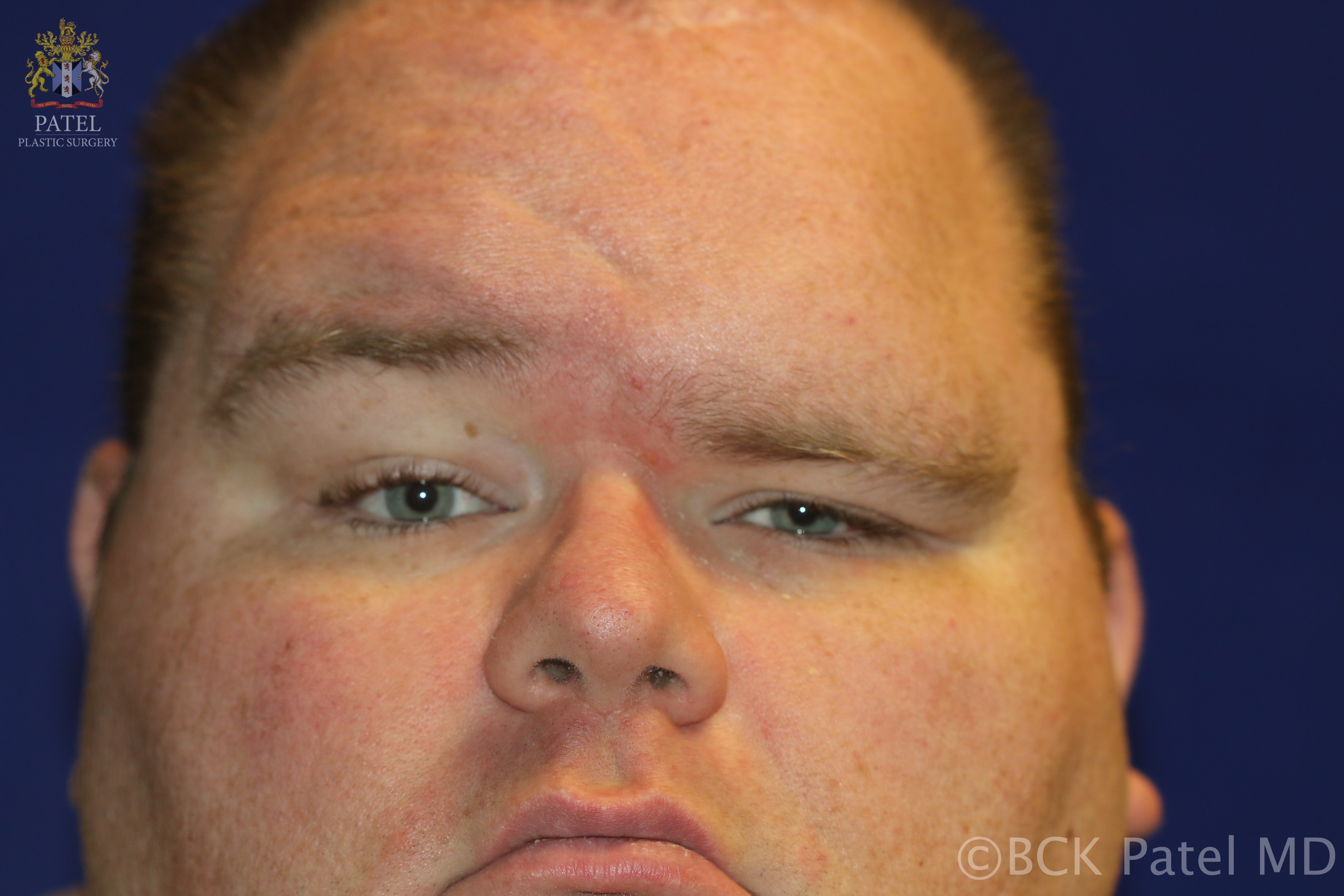
This 32-year-old male developed a left frontal branch of the facial nerve palsy after removal of an intracranial tumor 12 years ago. He has had three brow lift procedures which he claims all failed.
Contributed by Prof. Bhupendra C.K. Patel MD, FRCS