Flea Bites
- Article Author:
- Jackie Anderson
- Article Editor:
- Elizabeth Paterek
- Updated:
- 8/10/2020 10:52:55 PM
- For CME on this topic:
- Flea Bites CME
- PubMed Link:
- Flea Bites
Introduction
Siphonaptera, more commonly known as fleas, are an order of wingless arthropods with more than 2000 species. Though fleas survive in a multitude of environments, they are rare in low humidity environments and at elevations over 1500 ft.[1] Fleas survive as ectoparasites on warm-blooded species by consuming their blood. They are small, ranging in size from 1.0 to 3.3 mm, and have long hind legs adapted for jumping. Fleas are predominantly a nuisance to their host causing pruritic local reactions. They can also act as vectors for disease including typhus, rickettsial disease, bubonic plague, protozoan, and helminth infestations. Understanding these features can help to direct history taking, the physical exam, and treatment options. Additionally, exploring eradication techniques within the human environment can help to reduce the spread of disease from fleas to humans.
Etiology
Fleas typically bite when they come in contact with a host, commonly a cat or dog as well as other wildlife species including foxes and rats.[2][3] They feed off of the host’s blood and lay eggs in their fur. Ultimately, the flea’s eggs fall off of the animal allowing them to hatch in the host’s surrounding environment and perpetuating their lifecycle.
Epidemiology
Fleas are prevalent among mammals worldwide. In the developed world, the most influential vectors are household pets, specifically cats and dogs.[2] In the developing world, fleas are a significant cause of papular urticaria, especially in tropical regions; this is seen most commonly in pediatric patients with a frequency of 2.4 to 16.3%. While other insects can also cause a similar rash, fleas are one of the most frequent causes. In most patients, the rash improves by age 7, though in some cases it can last into adulthood.[4]
Tunga penetrans, also known as the chigoe or jigger flea, is a parasitic flea infestation found in tropical and subtropical climates including Africa, South America, and Central America. It is most common in underdeveloped and impoverished regions. Though rare in the United States and Europe despite global travel, it should be considered in patients returning from endemic regions. Peak incidence occurs during the dry season between August and September. Typically only adults are affected. Domestic and wild animals act as reservoirs of disease.[5]
Pathophysiology
Most flea bites result in papular urticaria, which is a dual Type I and Type IV hypersensitivity reaction. The reactions involve both IgE and cell-mediated responses. This rash appears as small, raised, erythematous, pruritic lesions. It most commonly affects exposed skin, generally, the feet and ankles and appears in clusters and linear patterns known as a "breakfast, lunch and dinner" pattern.[6][7]
Tungiasis is an inflammatory skin condition caused by T. penetrans which is the smallest known flea. Skin irritation occurs when a female flea embeds itself into the epithelium of the skin. Fleas typically embed themselves in soft areas of skin such as the medial foot, under nails and between toes. In cases of severe disease, the thigh, hands, perianal and groin may be involved. During the first 1 to 2 days of infestation, the host may notice itching or discomfort. The abdomen of the flea then swell with eggs forming a white pearly lesion. This increased pressure can cause a painful foreign body sensation and irritate surrounding neurovascular structures. Following this, the lesion becomes dark and crusted. It may create a honeycomb pattern in more extensive infection which is prone to secondary infection and increases the risk of gangrene and tetanus. After healing, scarring can occur.[5][8]
History and Physical
When evaluating a patient in whom there is a suspicion for flea bites, the provider should inquire about living conditions, recent exposures, and similar symptoms in co-habitants. The physical exam should include a thorough skin examination with particular attention to exposed areas such as the arms, legs, upper back, and scalp. Additionally, the exam should pursue details regarding any rashes including duration, edema, erythema, and pruritus. Evaluation for local areas of infection, while a rare presentation, should be performed as with all skin-breaks.
Evaluation
The diagnosis is typically from the history and physical exam. There are no laboratory tests that can confirm fleas as the causal agent of the rash. Confirmation of the diagnosis can instead be via examination of debris from vacuuming and bedding [9]. Flea bites typically result in papular urticaria, a rash that manifests as small, raised skin lesions. This reaction typically occurs within minutes of the bite and is characterized by local pruritus, swelling, and erythema. The ankles are the most common areas affected through any exposed skin such as the arms, legs, upper back, and scalp can show the rash. The buttocks, groin, genital, and axillary areas are typically spared. These bites may present in linear clusters referred to as a "breakfast, lunch and dinner" pattern.[10][11]
Treatment / Management
Skin subjected to flea bites, as with all insect bites, should be washed with soap and water. Local edema, erythema, and pruritus are symptoms of a hypersensitivity reaction caused by the bites and are treatable with cold packs and topical creams. Calamine or pramoxine-containing lotions and topical corticosteroids are effective at reducing local inflammation and pruritus. Oral antihistamines such as cetirizine or loratadine may also combat pruritus. Note that the application of topical antihistamines over large areas of skin should be avoided when oral H1 antihistamines are in use; such a combination can result in systemic anticholinergic toxicity. Oral steroids may be an option for severe swelling. The rash can take several weeks to resolve. Elimination of fleas from the patient’s surroundings is also imperative to stop the continuation of the rash.[9]
Tungarisis is usually benign and self-limited, especially in returning travelers. However, patients with extensive infestations may have complications including loss of nails, toe deformities and difficulty ambulating. Due to the high risk of secondary infection, patients with extensive infections should receive treatment with prophylactic antibiotics. In unvaccinated patients, tetanus risk is increased, and the practitioner should query all patients regarding their tetanus status. Conventional therapy is surgical extraction of the parasite under sterile conditions. No oral or topical therapy is entirely effective; however, in mild infections even without treatment the flea will die within 5 weeks and will be sloughed off with the skin. Basic hygiene as well as paving roads and floors can prevent infestation.[4][12][2]
Differential Diagnosis
The differential diagnosis of pruritic urticaria should focus on inciting factors such as recent changes in detergents and body products, as well as recent exposures to animals and insects. While fleas and mosquitoes are the most frequent insects associated, other insects can cause similar rashes.[4][12] Additionally, consideration should be given to the possibility of anaphylaxis by exploring the involvement of other organ systems including the gastrointestinal tract, as evidenced by vomiting and or diarrhea, and the pulmonary system, as evidenced by wheezing and or difficulty breathing. Anaphylaxis to insect bites, especially fleas, is very rare.
Prognosis
Flea bites typically have a benign clinical course. Most patients only experience a mild local reaction called papular urticaria. This rash resolves spontaneously and is manageable with anti-histamines and anti-inflammatory medications. Secondary infections such as cellulitis and abscess are possible and should have treatment per current clinical guidelines. Fleas can be vectors for secondary infections such as Yersinia pestis and murine typhus as well as parasitic infections. The prognosis for these patients is related to the secondary infection and not the flea bite.
Complications
Fleas are a critical factor in the epidemiologic cycles of several diseases including plague and typhus. As carriers of these diseases, they can spread them to humans through their bites.
Yersinia pestis, also known as the plague, is transmitted by fleas. Humans, after becoming infected with Y. pestis, are considered to be hosts but do not play an instrumental role in the natural disease cycle. In the United States, Y. pestis is most common in the south-western and Pacific coast states; however, the incidence of plague is low in the United States given that many of the regions affected are uninhabited.[13] While there are numerous routes of transmission of the plague to humans, flea bites remain the most common. Various mammalian species are carriers of the plague including cats, dogs, squirrels, mice, and rats.[14] Y. pestis, a gram-negative coccobacillus, manifests initially as regional lymphadenopathy secondary to an invasion of the lymphatic system. These inflamed lymph nodes are called buboes. Subsequent disease progression presents itself as pneumonia, hemorrhagic lesions, purpuric skin lesions, and sepsis secondary to bacteremia.[15] Treatment includes gentamycin and fluoroquinolones as first-line agents.
Murine typhus is transmitted by fleas, primarily the rat flea, Xenopsylla cheopis. It also spreads by the mouse flea, Leptopsyllia segnis, and the cat flea, Ctenocephalides. Inoculation of humans occurs through the contamination of bite wounds with the feces of infected fleas. In the United States, R. typhi highly correlates with areas with large rat populations. R. typhi and Rickettsia felis are also found to be maintained by domestic cats and opossums in suburban settings.[16] While Rickettsia felis does not cause murine typhus, the presentation of its infection is clinically indistinguishable from those of murine typhus. Manifestations include fever, headache, chills, myalgias, and other non-specific symptoms. A faint, maculopapular rash may also occur, spreading centrifugally but sparring the palms and soles.[17] Complications may occur in those with severe comorbidities including hepatic, cardiac, pulmonary, renal, and neurologic dysfunction.[17][18][19] Patient with uncomplicated typhus may receive treatment with doxycycline, which is more effective than azithromycin.[20] Chloramphenicol may be an option as a third-line agent.[21]
Deterrence and Patient Education
Prevention of flea bites involves avoiding exposure. Insecticidal sprays can be used in living conditions to eliminate fleas. Vacuuming and cleaning are also paramount because flea eggs drop onto floors after being laid in animal fur. Removing flea eggs from the environment halts the continuation of the flea's lifecycle.
Household pets, mostly cats and dogs, are common flea carriers. Flea preventatives are obtainable from a veterinarian. These agents can either topical or oral. Veterinary consultation is tantamount if household pets are suspected to be the source of the infestation.[22][23]
If in an environment where flea eradication is difficult or unfeasible, repellants are an option. Both DEET and thymol containing essential oils have demonstrated effectiveness against both human (Pulex irritans) and cat (Ctenocephalides felis) fleas.
Enhancing Healthcare Team Outcomes
Healthcare outcomes can be enhanced by open provider-patient communication. It is essential to an interprofessional team to ask about the patient’s living environment including contact with animals and provide tips on how to manage flea and other insect infestations. Assistance may be required from extermination services as well as veterinarians, in the case of pets in the home, to rid the environment of infestation. By counseling patients, the team members can help prevent both patient discomfort from bites as well as the spread of disease. Healthcare providers including nurse practitioners, nurses with specialty training in dermatology, pharmacists, and the primary care provider should provide a judgment-free space that offers a healthy exchange to educate the patient and family to facilitate the best patient care and produce the best outcomes. [Level V]
(Click Image to Enlarge)
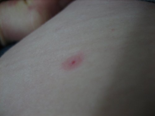
Flea bite with a hemorrhagic center and surrounding erythema.
Contributed from the Public Domain https://upload.wikimedia.org/wikipedia/en/7/78/Fleabite-closeup.jpg