Acrodermatitis Papular
- Article Author:
- Jessica Snowden
- Article Author:
- Ashley Rice
- Article Editor:
- Noreen O'Shea
- Updated:
- 7/14/2020 9:22:17 AM
- For CME on this topic:
- Acrodermatitis Papular CME
- PubMed Link:
- Acrodermatitis Papular
Introduction
Papular acrodermatitis of childhood, otherwise known as Gianotti-Crosti syndrome, is a benign rash that is associated with a wide variety of viral illnesses. While historically thought to be solely a manifestation of hepatitis B infection, it has been demonstrated to occur following many viral illnesses and vaccination, which suggests that this is an immunologic response rather than a primary manifestation of infection. Papular acrodermatitis of childhood is characterized by the acute eruption of monomorphic skin-colored to pink-red papules on the face, buttocks, and extensor surfaces of the extremities. The lesions usually spontaneously resolve and treatment is supportive.[1][2][3]
Etiology
When papular acrodermatitis of childhood was initially described in the 1950s, it was believed to be a manifestation of hepatitis B virus infection. This was in large part due to the prevalence of anicteric hepatitis in these patients. While this association is still common in hepatitis B endemic areas, it is rare in the United States since the advent of the hepatitis B vaccine. Ongoing studies have demonstrated that papular acrodermatitis of childhood can occur following a wide variety of viruses. These viruses include but are not limited to Epstein-Barr virus, cytomegalovirus, coxsackievirus, adenovirus, influenza, enteroviruses, echovirus, hepatitis A virus, herpes simplex viruses, HHV-6, HIV, mumps, parainfluenza virus, parvovirus B19, poxviruses, respiratory syncytial virus, and rotavirus. Additionally, it has been reported after vaccination, including influenza, Calmette-Guerin bacillus, diphtheria-pertussis-tetanus, poliomyelitis, hepatitis B, Japanese encephalitis, and measles vaccines. The Epstein-Barr virus is the most commonly reported cause of papular acrodermatitis of childhood in the United States. However, in many cases, no infectious trigger is identified. The relationship between these potential triggers and the manifestation of papular acrodermatitis of childhood is not well defined but is presumed to be immunologically mediated.[4][5][6][7][8][9]
Epidemiology
Reports of papular acrodermatitis of childhood are most common in children younger than four years of age, which is consistent with its viral etiology. In children, there is no sex predilection; however, in adulthood, it is slightly more common in female patients. In papular acrodermatitis of childhood, there does not appear to be a genetic or familial predisposition. Most cases occur in spring and summer. Papular acrodermatitis of childhood occurs more often in children with atopic diseases, including atopic dermatitis. Although the disease is observed worldwide, the etiological agents exhibit geographic variations.[7][10][11]
Pathophysiology
The precise pathophysiology of papular acrodermatitis is unknown, but it is presumed to be immunologically mediated given its association with viral infections and vaccination. Its increased prevalence in patients with a history of atopic disease, family history of atopic disease, and/or elevated immunoglobulin E also supports an immune-mediated pathology. Proposed processes include immune complexes or delayed hypersensitivity reactions, but to date, this has not been well defined.
Histopathology
The histopathology of papular acrodermatitis of childhood is non-specific. Findings may include parakeratosis and focal, epidermal spongiosis. A perivascular, lymphocytic infiltrate may also be present within the papillary dermis.[12]
History and Physical
As its name implies, papular acrodermatitis of childhood clinically manifests as an acute, symmetric eruption of flat-topped papules in an acral distribution. These monomorphic papules most commonly occur on the extensor surfaces of the extremities. Arms are more commonly involved than legs. The face, buttocks, palms, and soles may also be affected. The trunk and scalp are relatively spared, as are the popliteal and antecubital fossae. However, the involvement of any of these areas does not preclude the diagnosis of papular acrodermatitis of childhood. The lesions are usually pale pink to flesh-colored and 1 mm to 10 mm in size. The lesions tend to be larger in younger children and smaller in older children and adolescents. Rarely, lesions may be vesicular or hemorrhagic. They are firm and discrete. Rarely, the lesions can become confluent over pressure points like knees and elbows. Koebner phenomena, the accentuation of lesions at sites of trauma, has also been reported. Mild-to-moderate pruritus may be present. Symptoms frequently last 2 to 4 weeks. In some cases, new lesions may continue to appear for up to 8 to 11 weeks after onset of illness. Importantly, the appearance of the rash itself may not help to distinguish the variously associated etiologies of papular acrodermatitis of childhood; however, careful assessment of associated or antecedent symptoms is essential for recognizing possible infectious triggers that may require further evaluation or isolation. Usually, the rash resolves without any residual effects or scarring. Occasionally, hyperpigmentation or hypopigmentation may persist after the rash resolves.[7][13][14][15] There is no associated mucosal enanthem but mucocutaneous features of the underlying viral trigger may be present.[15]
Fever, lymphadenitis, and other symptoms such as hepatosplenomegaly or pharyngitis may be seen in association with the rash or preceding it. This is consistent with a variety of viral triggers for this entity. There may also be a history of vaccination preceding the onset of the rash.[13][16] There are no specific laboratory findings seen in papular acrodermatitis of childhood. However, modest lymphocytosis or lymphopenia and elevated liver enzymes may be seen in Epstein-Barr virus, cytomegalovirus, or hepatitis-associated disease.[13]
Evaluation
In general, papular acrodermatitis is a clinical diagnosis with no laboratory testing indicated unless there are symptoms or physical exam findings, such as hepatomegaly, to suggest an underlying viral illness, such as hepatitis, that may require further management. Diagnostic criteria have been proposed and include the following symptoms:
- Lesions lasting at least 10 days;
- 1 mm to 10 mm papules or papulovesicular lesions with involvement of three of the four following sites: extensor surfaces of the forearms, extensor surfaces of the legs, cheeks, or buttocks;
- Symmetrical distribution of lesions.
Practitioners may need to further evaluate the causative virus in children who are immunocompromised or who reside in endemic areas. If a patient presents with jaundice, hepatomegaly, or generalized lymphadenopathy, evaluation for other viral infections, including hepatitis B or Epstein-Barr virus, may be indicated. Rarely, a skin biopsy may be performed to exclude other conditions in immunocompromised or other high-risk patients. Normally, biopsy findings are non-specific, with dermal perivascular mononuclear cell infiltrates and capillary endothelial swelling.[17][18]
Treatment / Management
Practitioners should reassure patients that papular acrodermatitis on its own is a benign and self-limited illness. The majority of cases spontaneously resolve without active intervention. Symptomatic management of itching may be helpful. Patients may use emollients or oral antihistamines. Since infectious agents do not directly cause skin lesions, mild to moderate potency topical steroids may be used in patients who fail to achieve relief of itching with emollients and oral antihistamines. However, treatment with topical steroids or oral antihistamines does not shorten the course of illness. Any underlying disorder, such as hepatitis B, that requires further management should be addressed. Lesions resolve without scarring although post-inflammatory pigmentary alteration may persist in some patients.[19]
Differential Diagnosis
The differential diagnosis of papular acrodermatitis includes other viral exanthems such as erythema infectiosum and Hand, Foot, and Mouth disease, insect bites, scabies, papular urticaria, atopic dermatitis, erythema multiforme, Langerhans cell histiocytosis, lichenoid dermatoses, and IgA vasculitis.
Prognosis
Papular acrodermatitis of childhood is a benign and self-limited disease that typically persists for 10 days to 6 months. The majority of skin manifestations resolve within 2 weeks to 2 months. Extracutaneous manifestations tend to persist longer than cutaneous lesions. Recurrences are rare.[16][20][21]
Complications
Deterrence and Patient Education
It is important to reassure parents about the benign nature of papular acrodermatitis of childhood to avoid unnecessary invasive testing or treatments. Further laboratory work-up is only indicated in high-risk patients, including those who are immunocompromised or from hepatitis B endemic areas. Additional testing is also suggested in those with extracutaneous symptoms suggestive of an underlying viral etiology such as Epstein-Barr virus.
It is not recommended to exclude children from daycare, school, or social activities.
Enhancing Healthcare Team Outcomes
Healthcare workers, including nurse practitioners and clinicians, need to be aware that papular acrodermatitis of childhood is a benign viral-induced skin disorder. It is often confused with other skin infections of childhood. If there is any doubt about the diagnosis, the patient should be referred to a dermatologist. Papular acrodermatitis of childhood is not person-to-person transmissible and does not require isolation beyond standard precautions or restrictions from school or other community activities. However, the associated viral infection may be contagious, thus isolation may need to be considered based on other symptoms suggestive of infectious disease. The dermatologist and dermatologist nurse specialist should provide a team effort to educate patients and their families about the disease and the necessary precautions. [Level V]
(Click Image to Enlarge)
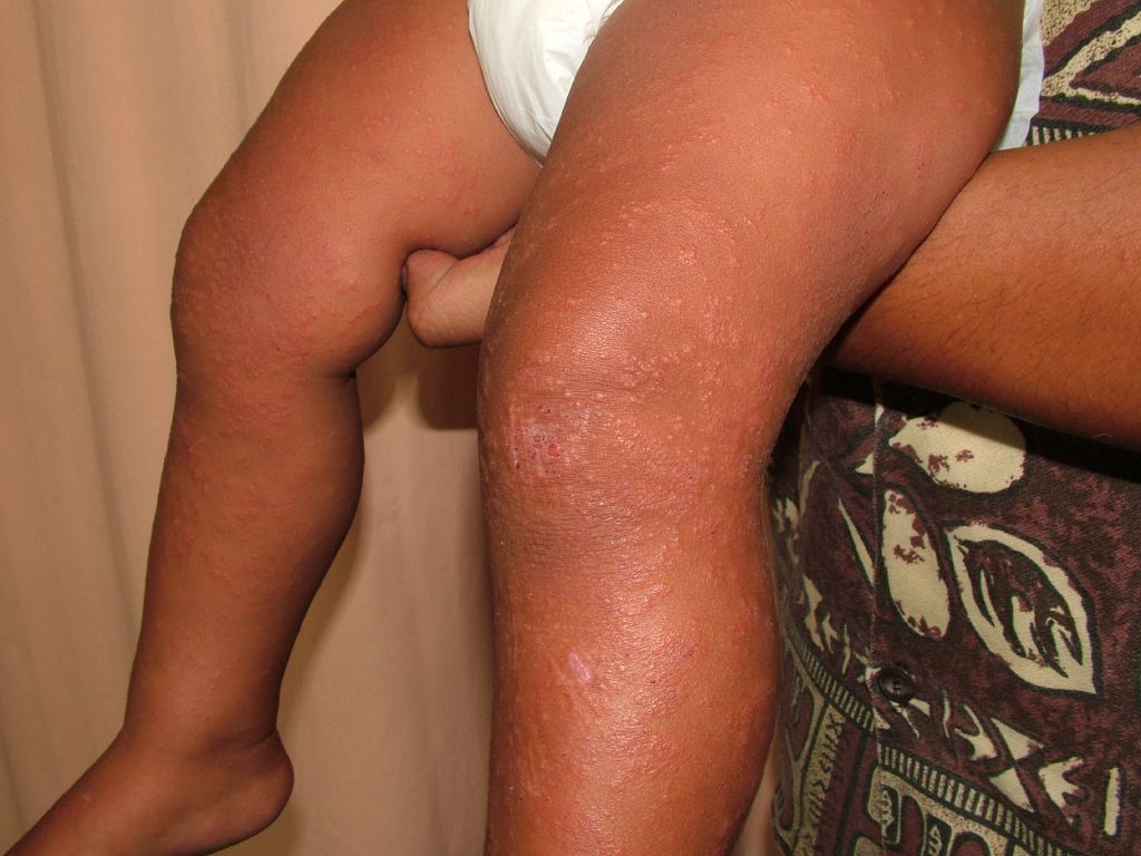
Gianotti Crosti
Contributed by DermNetNZ